. 2015 Nov 15;195(10):4884-91.
doi: 10.4049/jimmunol.1501194. Epub 2015 Oct 2.

Preferential Destruction of Interstitial Macrophages over Alveolar Macrophages as a Cause of Pulmonary Disease in Simian Immunodeficiency Virus-Infected Rhesus Macaques

Affiliations

Preferential Destruction of Interstitial Macrophages over Alveolar Macrophages as a Cause of Pulmonary Disease in Simian Immunodeficiency Virus-Infected Rhesus Macaques

Yanhui Cai et al. J Immunol. .

Abstract

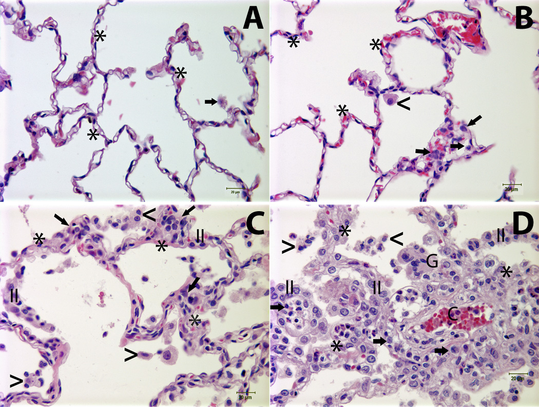
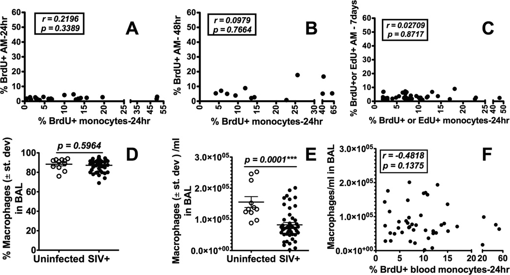
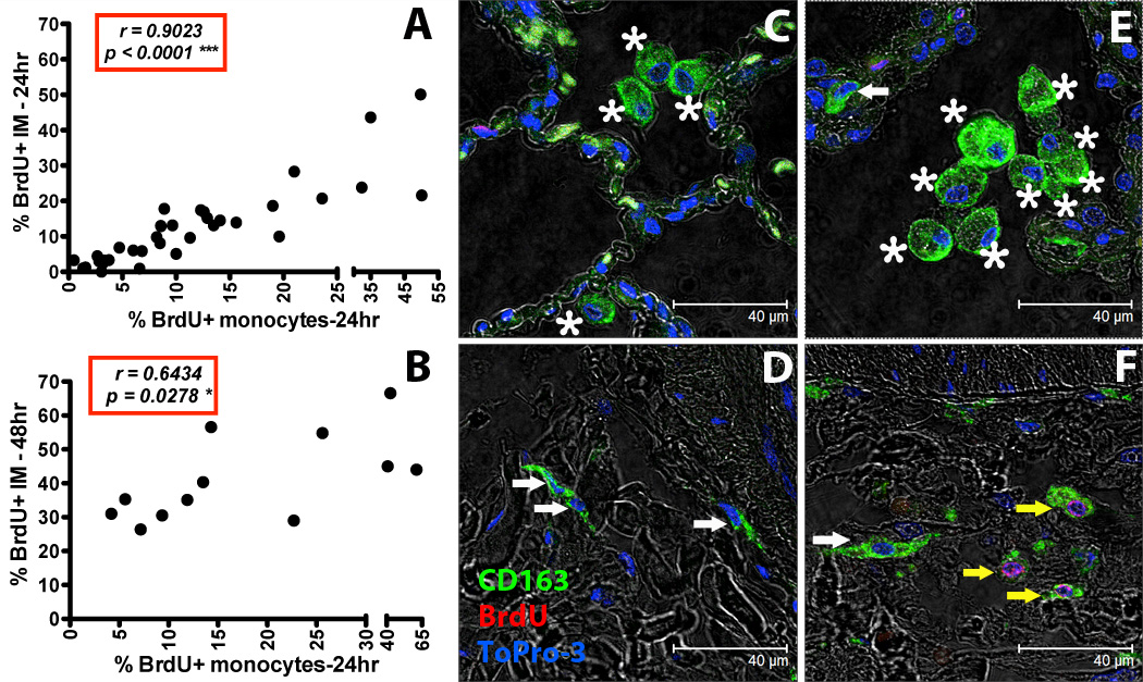
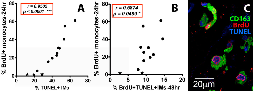
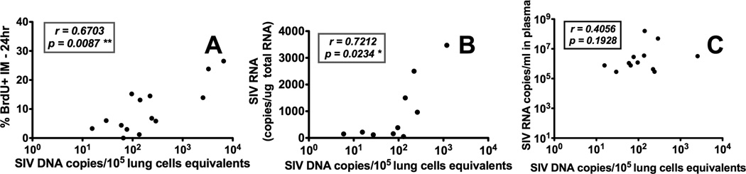
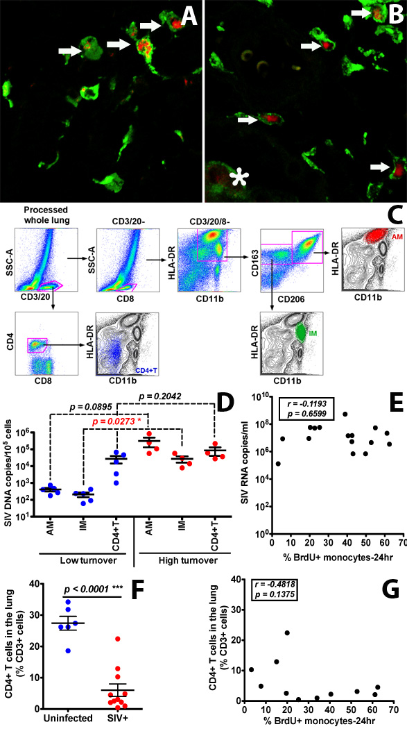
To our knowledge, this study demonstrates for the first time that the AIDS virus differentially impacts two distinct subsets of lung macrophages. The predominant macrophages harvested by bronchoalveolar lavage (BAL), alveolar macrophages (AMs), are routinely used in studies on human lung macrophages, are long-lived cells, and exhibit low turnover. Interstitial macrophages (IMs) inhabit the lung tissue, are not recovered with BAL, are shorter-lived, and exhibit higher baseline turnover rates distinct from AMs. We examined the effects of SIV infection on AMs in BAL fluid and IMs in lung tissue of rhesus macaques. SIV infection produced massive cell death of IMs that contributed to lung tissue damage. Conversely, SIV infection induced minimal cell death of AMs, and these cells maintained the lower turnover rate throughout the duration of infection. This indicates that SIV produces lung tissue damage through destruction of IMs, whereas the longer-lived AMs may serve as a virus reservoir to facilitate HIV persistence.


Figures

Figure 1
Figure 1. The severity of pulmonary lesions correlated with the rate of monocyte turnover (RMT) in SIV-infected rhesus macaques
Panel A: Normal lung tissue from an uninfected monkey [the rate of monocyte turnover (RMT) = 1.61%]; Panel B: Lung tissue from an SIV-infected animal (RMT = 22.7%) demonstrates minimal interstitial accumulation of a few mononuclear cells (arrows) and rare alveolar macrophages (<); Panels C and D: Lung tissues from SIV-infected animals with RMT = 40.5% and RMT= 55%, respectively, exhibited mild (Panel C) to moderate (Panel D) interstitial pneumonia characterized by alveolar septa thickening, capillary dilation (C), increased the numbers of mononuclear cells (arrows) and hyperplastic type II pneumocytes (II). Low (Panel C) to moderate (Panel D) numbers of alveolar macrophages (<) and multinucleated giant cells (G) were noted in the alveolar spaces. Asterisk (*) = alveolar septum. Tissues were stained with H&E and viewed at 400× magnification. Lung injury was assessed using a histological scoring system as previously described (4, 16).
Figure 2
Figure 2. Increased turnover of blood monocytes in SIV-infected macaques is not reflected in the BAL
BAL samples were collected prior to and during different stages of SIV infection from rhesus macaques. BrdU or EdU was injected i.v. into SIV-infected rhesus macaques and the turnover of blood monocytes and AM recovered from BAL was analyzed by flow cytometry. There was no statistically significant correlation between blood monocyte turnover and AM turnover at 24 hr after BrdU/EdU injection (A: n= 21), 48 hr (B: n=12) and 7 days later (C: n=38) using Spearman’s correlation analysis. There also was no difference in the percent of macrophages recovered in BAL from SIV-infected vs uninfected macaques (D) although there was a significant decrease in the absolute number of macrophages recovered from BAL of SIV-infected compared to uninfected macaques by Student’s t test (E). The numbers of macrophages recovered from BAL of SIV-infected monkeys did not correlate with blood monocyte turnover by Spearman’s correlation analysis (F).
Figure 3
Figure 3. Increased turnover rate of lung IM correlates with increased blood monocyte turnover in SIV-infected macaques
BrdU or EdU was injected i.v. into SIV-infected or uninfected rhesus macaques and lung IM were analyzed by flow cytometry (A and B). By Spearman’s correlation analyses, a significant correlation was observed between blood monocyte turnover and IM turnover 24 hr (A: n = 35) or 48hr (B: n = 12) after BrdU/EdU injection. Paraffin-embedded lung tissue sections obtained after necropsy from uninfected (C and D: representative of 4 animals) and SIV-infected rhesus macaques (E and F: representative of 4 animals) were stained with anti-CD163 antibody (macrophages-Green), anti-BrdU antibody (turnover-Red) and Topro-3 (nucleic acid-Blue). Images were captured with a Leica TCS SP2 confocal microscope equipped with a 3-laser (Leica Microsystems) under an oil objective (63×, fluotar/NA 1.0) for a final magnification of 1260×. White arrows indicate CD163-staining IM cells (green), yellow arrows indicate CD163-staining IM cells that incorporated BrdU (green and red), and asterisks indicate CD163-staining AM (green).
Figure 4
Figure 4. Increased death rate of lung IM correlates with increased blood monocyte turnover rate in SIV-infected rhesus macaques
(A) Spearman’s correlation analyses were performed to relate blood monocyte turnover (BrdU) and percent of TUNEL+ IM (cell death) from lungs of uninfected and SIV-infected monkeys collected 24 hr post BrdU injection (n=14), and (B) 48 hr post BrdU injection (n=12). A minimum of 400 cells were counted in at least 5 microscopic fields of lung tissues from SIV-infected (●) and uninfected (★) monkeys. (C) A representative image of IM in the lung tissues from an SIV-infected monkey with high monocyte turnover (55% BrdU-staining monocytes at 24h) is shown. Lung tissues from four uninfected rhesus macaques and ten SIV-infected monkeys with different monocyte turnover rates were stained with anti-CD163 (Green) and anti-BrdU (turnover-Red) antibodies. Cell death/apoptosis was measured by TUNEL (Blue) staining. Images were captured with a Leica TCS SP2 confocal microscope at a final magnification of 1260×.
Figure 5
Figure 5. Increased SIV levels in lung tissues of infected rhesus macaques correlate with increased IM and blood monocyte turnover rates but not with SIV plasma viral loads (pVL)
Lung tissues were collected from SIV-infected macaques at different stages of disease and quantitated for SIV DNA and RNA. (A) The number of SIV DNA copy equivalents in lung tissue directly correlated with IM turnover in SIV-infected macaques (n=14). (B) SIV DNA copy numbers directly correlated with SIV RNA copy numbers in the lungs of SIV-infected macaques (n=10). (C) SIV DNA copy numbers in lung did not correlate with the pVL in SIV-infected macaques (n=12). Spearman’s correlation analyses were applied for statistical analysis.
Figure 6
Figure 6. Infection and replication of SIV in IM and AM contribute to the viral load in lung tissues of infected rhesus macaques exhibiting high monocyte turnover
(A&B) Confocal microscopy was performed on lung tissues obtained from SIV-infected monkeys with low (≤ 30%) monocyte turnover (A: n=3) and higher (> 30%) blood monocyte turnover levels (B: n=6). Anti-CD163 (Green) antibody was used to identify macrophages, and SIV RNA (Red) was detected with anti-sense riboprobes. Arrows indicate SIV-infected IM. The asterisk indicates an SIV-infected AM. (C) Gating strategy for analyzing and sorting AM, IM and CD4+ T cells isolated from lung tissue. (D) IM, AM and CD4+ T cells were sorted via FACS from single-cell suspensions of lung tissues from SIV-infected monkeys with low (n=5) and high (n=4) blood monocyte turnover, and SIV DNA levels were quantitated and standardized against RNase P levels. (E) Plasma VL did not correlate with monocyte turnover in SIV-infected macaques. (F) CD4+ T cell depletion in the lung of all SIV-infected macaques was evident (p<0.0001). (G) The levels of CD4+ T cells did not correlate with monocyte turnover rates in SIV-infected macaques. Student’s t test was applied for comparisons in results shown in panels D and F, and Spearman’s correlation analysis was applied in results shown in panels E and G.

References

    1. Van Rompay KK. The use of nonhuman primate models of HIV infection for the evaluation of antiviral strategies. AIDS research and human retroviruses. 2012;28:16–35. - PubMed
    1. Hasegawa A, Liu H, Ling B, Borda JT, Alvarez X, Sugimoto C, Vinet-Oliphant H, Kim WK, Williams KC, Ribeiro RM, Lackner AA, Veazey RS, Kuroda MJ. The level of monocyte turnover predicts disease progression in the macaque model of AIDS. Blood. 2009;114:2917–2925. - PMC - PubMed
    1. Burdo TH, Soulas C, Orzechowski K, Button J, Krishnan A, Sugimoto C, Alvarez X, Kuroda MJ, Williams KC. Increased monocyte turnover from bone marrow correlates with severity of SIV encephalitis and CD163 levels in plasma. PLoS pathogens. 2010;6:e1000842. - PMC - PubMed
    1. Cai Y, Sugimoto C, Liu DX, Midkiff CC, Alvarez X, Lackner AA, Kim WK, Didier ES, Kuroda MJ. Increased monocyte turnover is associated with interstitial macrophage accumulation and pulmonary tissue damage in SIV-infected rhesus macaques. Journal of leukocyte biology. 2015;97:1147–1153. - PMC - PubMed
    1. Lin SL, Castano AP, Nowlin BT, Lupher ML, Jr, Duffield JS. Bone marrow Ly6Chigh monocytes are selectively recruited to injured kidney and differentiate into functionally distinct populations. J Immunol. 2009;183:6733–6743. - PubMed

Publication types

MeSH terms