. 2016 Jan 1;370(1):1-9.
doi: 10.1016/j.canlet.2015.09.016. Epub 2015 Oct 1.

Activation of signal transduction pathways during hepatic oncogenesis

Affiliations

Activation of signal transduction pathways during hepatic oncogenesis

Waihong Chung et al. Cancer Lett. .

Abstract

Background and aims: Understanding the molecular pathogenesis of hepatocellular carcinoma (HCC) is essential to identify therapeutic targets. A hepatitis B virus (HBV) related double transgenic murine model was developed.

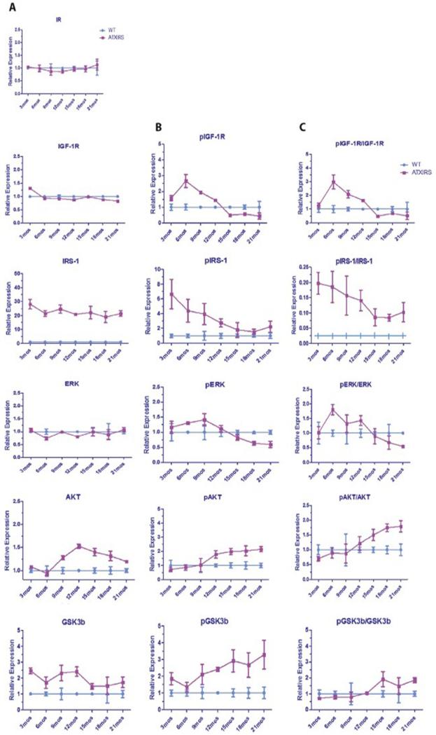
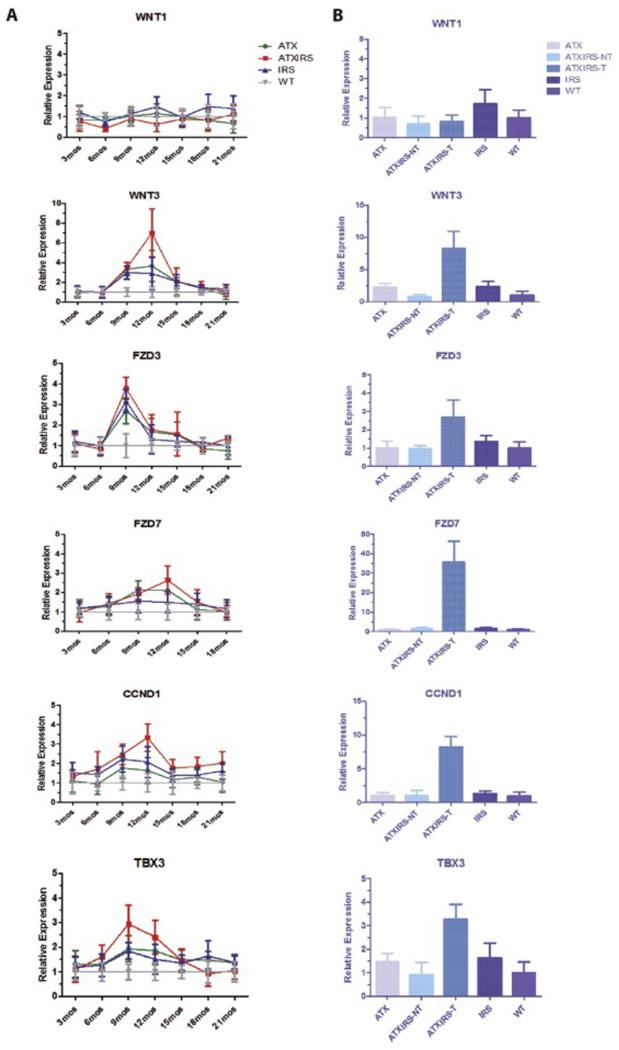
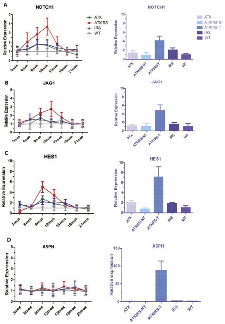
Methods: Liver specific expression of HBV X protein (HBx) and insulin receptor substrate 1 (IRS1) was achieved and transgenic mice were followed from birth to age 21 months. Liver and tumor tissue were assessed for histologic changes as well as activation of signal transduction pathways by qRT-PCR and multiplex ELISA protein assays.

Results: Overexpression of HBx and IRS1 stimulates liver cell proliferation in the double transgenic mice. Only the male mice developed HCC starting at age 15-18 months. The IN/IGF1/IRS1/MAPK/ERK and IN/IGF1/IRS1/PI3K/AKT/GSK3β cascades were activated early (6-9 months) in the liver followed by WNT/β-catenin and Notch signaling. Aspartate β-hydroxylase (ASPH) was found to link these upstream growth factor signaling pathways to downstream Notch activation in tumor tissues.

Conclusions: Sustained overexpression of HBx and IRS1 led to constitutive activation of a tripartite growth factor signal transduction cascade in the liver and was necessary and sufficient to promote HCC development and progression.

Keywords: Growth factor signaling pathways; Hepatocellular carcinoma; Transgenic mice.


Figures

Figure 1
Figure 1
Phenotypic features of tumor development in the ATX/IRS1 double transgenic mice. (A) Proliferative stimulus provided by expression of the HBx and IRS1 transgenes. Measurement of PCNA expression by qRT-PCR in WT, ATX, IRS1, and ATX/IRS1 livers. Note the comparison to the PCNA level found in ATX/IRS1 generated HCC tumors compared to WT (p=0.00005). Incidence of HCC development in female and male ATX/IRS1 transgenic mice. In males tumor development and progression starts after 15 months of age. (B) Morphologic appearance of HCC found in ATX/IRS1 male transgenic mice at 18 months of age. (C) Histologic features of HCC where approximately 70% of tumors was surrounded by a fibrous capsule (arrows).
Figure 2
Figure 2
Serial changes in IN/IGF1/IRS1/RAS/RAF/MAPK/ERK and IN/IGF1/IRS1/PI3K/AKT/GSK3β signaling cascades in the livers of male WT vs. ATX/IRS1 double transgenic mice as measured by Multiplex ELISA assays. (A, B and C) Left panel represents total protein expression, middle panel represents phosphorylated proteins and right panel represents the ratio of protein/phosphoprotein serially studied from 3 to 21 months of age. Insulin receptor (IR) showing no change in expression. Early (3 month) increase in pIGF1R ratio compared to WT (p<0.003). Enhanced phosphorylation of IRS1 in transgenic liver at 3 months compared to WT (p<0.0001). Early (3-6 months) enhanced phosphorylation of ERK (p<0.01). Elevated expression and phosphorylation of AKT at 9-12 month of age and persistence from 12-21 months (p<0.003). Enhanced expression and phosphorylation of GSK3β that persisted for 21 months compared to WT (p<0.001). The findings suggest that activation of these two growth factor signaling pathways occur early during the spontaneous development and growth of HCC due to constitutive overexpression of HBx and IRS1 transgenes and they persist in the liver up to 21 months of age (characterized by pAKT and pGSK3β).
Figure 3
Figure 3
Serial studies in single and double transgenic mice liver demonstrating activation of the WNT/β-catenin pathway during tumorigenesis. The left panel (A) represents RT-PCR results in the liver from different time points and the right panel (B), a direct comparison to HCC tumor tissue. WNT1 expression was minimally elevated (p>0.05) in IRS1 transgenic liver at 18 months. WNT3 expression was highly upregulated in ATX/IRS1, to a less extent in ATX or IRS1 single transgenic mice at 12 months as compared to WT (p<0.0001, p<0.001, p<0.01) respectively. Tumor tissue exhibited a high level WNT3 expression in single and double transgenic mice, compared to liver tissue derived from WT (p<0.00005). FZD3 expression was substantially upregulated in transgenic mice at 9 months compared to WT and overexpressed in tumor tissue as well (p<0.008). FZD7 was overexpressed at 9-12 months in transgenic liver (p<0.0006) and strikingly upregulated in tumor tissue (p<0.0004). The downstream WNT/β-catenin regulated gene (cyclin D1) was upregulated between 9-12 months (p<0.001) and highly overexpressed in tumor tissue (p<0.00007). The transcription factor TBX3 as a representative downstream target of WNT/β-catenin was substantially upregulated in the ATX/IRS1 mice (p<0.003) as well as tumors derived from these animals (p<0.001).
Figure 4
Figure 4
Serial studies on Notch activation during hepatic oncogenesis in transgenic mice liver at different time points and among different genotypes as measured by qRT-PCR. (A) Upregulation of Notch1 gene expression in ATX/IRS1 mice that peaked at 12 months (p<0.009) with overexpression in tumor tissue (p<0.00001). (B) Enhanced expression of the Jag1 ligand both in the liver of double transgenic male mice (p<0.01) and their derived HCC tumors (p<0.00003). (C) Downstream Notch regulated Hes1 gene was also upregulated (9 months) in the ATX/IRS1 transgenic male mice (p<0.0007) and tumor tissue (p<0.00004). (D) ASPH levels were unchanged in the liver of WT and transgenic animals but was elevated 70 fold in HCC tumors (p<0.00001).
Figure 5
Figure 5
A. Crosstalk of HBx with IN/IGF and Wnt/β-catenin signaling cascades. HBx can upregulate IN/IGF signaling by activating Ras and upregulate Wnt/β-catenin signaling by suppressing GSK3β. An extensive network of crosstalk and feedback circuit exists between the two pathways, the PI3K/AKT cascade downstream of IN/IGF can suppress GSK3β activity through an inhibitory phosphorylation event. B. Hypothesized network of pathways/molecules involved in HCC oncogenesis in ATX/IRS1 transgenic mice. ASPH acts as a link between the IN/IGF growth factors and the downstream pathways, especially activation of Notch signaling. ASPH expression can be upregulated by IN or IGF1/2 stimulation leading to activation of Notch to promote cell migration, invasion and metastasis of HCC. A small molecule inhibitor (SMI) that inhibits β-hydroxylase activity by 80% demonstrates anti-tumor effects on HCC growth and progression [5].

References

    1. Hollinger FB, Purcell RH, Gerin JL, Ganem DE, Feinstone SM. Viral Hepatitis. 1st Edition Lippincott Williams & Wilkins; 2002.
    1. Schluter V, Meyer M, Hofschneider PH, Koshy R, Caselmann WH. Integrated hepatitis B virus X and 3′ truncated preS/S sequences derived from human hepatomas encode functionally active transactivators. Oncogene. 1994;9:3335–3344. - PubMed
    1. Nishiyama M, Wands JR. Cloning and increased expression of an insulin receptor substrate-1-like gene in human hepatocellular carcinoma. Biochem Biophys Res Commun. 1992;183:280–285. - PubMed
    1. Longato L, de la Monte S, Kuzushita N, Horimoto M, Rogers AB, Slagle BL, Wands JR. Overexpression of insulin receptor substrate-1 and hepatitis Bx genes causes premalignant alterations in the liver. Hepatology. 2009;49:1935–1943. - PMC - PubMed
    1. Aihara A, Huang CK, Olsen MJ, Lin Q, Chung W, Tang Q, Dong X, Wands JR. A Cell surface beta-Hydroxylase is a biomarker and therapeutic target for hepatocellular carcinoma. Hepatology. 2014 - PMC - PubMed

Publication types