Review
. 2016 Jan;73(1):79-94.
doi: 10.1007/s00018-015-2052-6. Epub 2015 Oct 3.

The endoplasmic reticulum: structure, function and response to cellular signaling

Affiliations
Review

The endoplasmic reticulum: structure, function and response to cellular signaling

Dianne S Schwarz et al. Cell Mol Life Sci. 2016 Jan.

Abstract

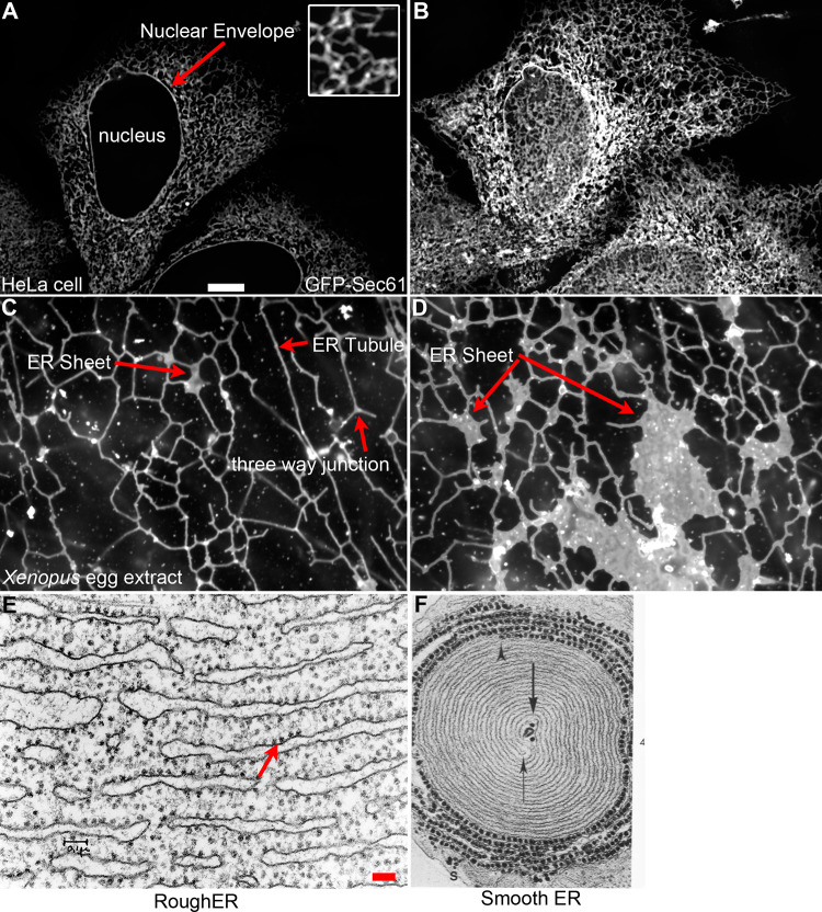
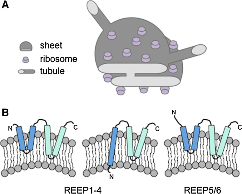
The endoplasmic reticulum (ER) is a large, dynamic structure that serves many roles in the cell including calcium storage, protein synthesis and lipid metabolism. The diverse functions of the ER are performed by distinct domains; consisting of tubules, sheets and the nuclear envelope. Several proteins that contribute to the overall architecture and dynamics of the ER have been identified, but many questions remain as to how the ER changes shape in response to cellular cues, cell type, cell cycle state and during development of the organism. Here we discuss what is known about the dynamics of the ER, what questions remain, and how coordinated responses add to the layers of regulation in this dynamic organelle.

Keywords: Fertilization; Interphase; Mitosis; Organization; Phosphorylation; Unfolded protein response.


Figures

Fig. 1
Fig. 1
Various ER structural morphologies. a Location of the ER visualized in a HeLa cell transfected with GFP-Sec61β. Inset shows the polygonal network of the peripheral ER magnified ×3 relative to the magnification in a. This view highlights the relationship of the ER to the nuclear envelope (red arrow). b ER morphology from the same HeLa cell depicting an image plane closer to the coverslip. This highlights the complexity of the peripheral ER. c ER network formed in Xenopus egg extracts. Three-way junctions, ER tubules and small ER sheets are highlighted (red arrows). d ER network formed in Xenopus egg extracts highlighting large ER sheets containing ribosomes (red arrow). Scale bar for ad is 10 μM and is shown in a. e Electron micrograph (EM) of rough ER from guinea pig pancreas. Reprinted with permission from James Jamieson. Scale bar is 0.1 μM. f EM of smooth ER from ocular rabbit muscle. Reprinted with permission from Fig. 4 [164]. Magnification is ×50,000
Fig. 2
Fig. 2
Structure of ER sheets and tubules. a ER sheets and tubules have a diameter of 30–50 nm in eukaryotes. Eukaryotic ribosomes are 25–30 nm and localize to the flat regions of ER sheets, giving the sheets a rough appearance (rough ER). Ribosomes are present in much lower numbers on tubules, giving the tubules a more smooth appearance (smooth ER). b Models of potential hairpin topologies of REEP family proteins that act as wedges to promote bending of the membrane, adapted from [63]

References

    1. Reid DW, Nicchitta CV. Diversity and selectivity in mRNA translation on the endoplasmic reticulum. Nat Rev Mol Cell Biol. 2015;16(4):221–231. - PMC - PubMed
    1. Rapoport TA. Protein translocation across the eukaryotic endoplasmic reticulum and bacterial plasma membranes. Nature. 2007;450(7170):663–669. doi: 10.1038/nature06384. - DOI - PubMed
    1. Braakman I, Hebert DN. Protein folding in the endoplasmic reticulum. Cold Spring Harb Perspect Biol. 2013;5(5):a013201. doi: 10.1101/cshperspect.a013201. - DOI - PMC - PubMed
    1. Fagone P, Jackowski S. Membrane phospholipid synthesis and endoplasmic reticulum function. J Lipid Res. 2009;50(Suppl):S311–S316. - PMC - PubMed
    1. Hebert DN, Garman SC, Molinari M. The glycan code of the endoplasmic reticulum: asparagine-linked carbohydrates as protein maturation and quality-control tags. Trends Cell Biol. 2005;15(7):364–370. doi: 10.1016/j.tcb.2005.05.007. - DOI - PubMed