. 2015 Oct 5:5:14603.
doi: 10.1038/srep14603.

Inflammation-associated alterations to the intestinal microbiota reduce colonization resistance against non-typhoidal Salmonella during concurrent malaria parasite infection

Affiliations

Inflammation-associated alterations to the intestinal microbiota reduce colonization resistance against non-typhoidal Salmonella during concurrent malaria parasite infection

Jason P Mooney et al. Sci Rep. .

Abstract

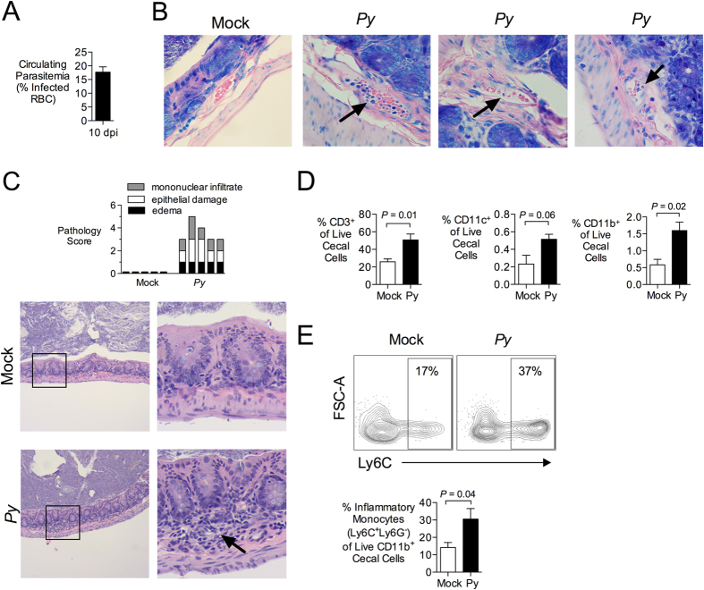
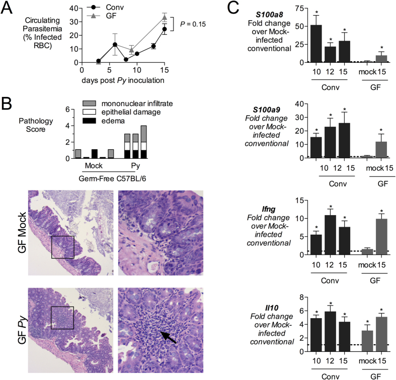
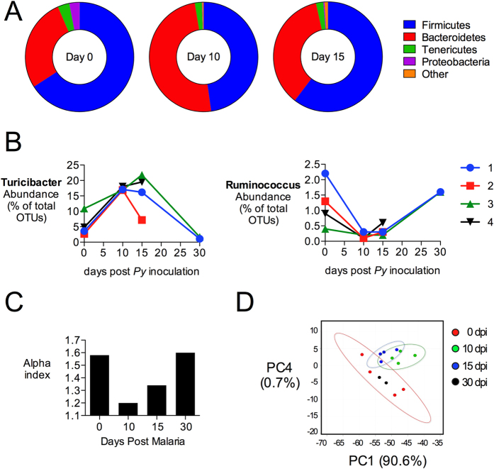
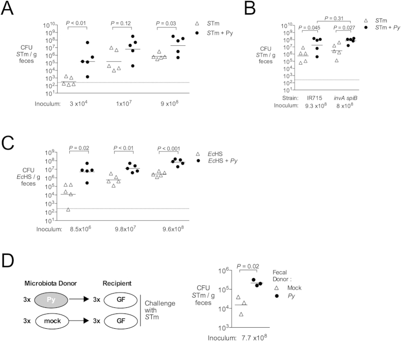
Childhood malaria is a risk factor for disseminated infections with non-typhoidal Salmonella (NTS) in sub-Saharan Africa. While hemolytic anemia and an altered cytokine environment have been implicated in increased susceptibility to NTS, it is not known whether malaria affects resistance to intestinal colonization with NTS. To address this question, we utilized a murine model of co-infection. Infection of mice with Plasmodium yoelii elicited infiltration of inflammatory macrophages and T cells into the intestinal mucosa and increased expression of inflammatory cytokines. These mucosal responses were also observed in germ-free mice, showing that they are independent of the resident microbiota. Remarkably, P. yoelii infection reduced colonization resistance of mice against S. enterica serotype Typhimurium. Further, 16S rRNA sequence analysis of the intestinal microbiota revealed marked changes in the community structure. Shifts in the microbiota increased susceptibility to intestinal colonization by S. Typhimurium, as demonstrated by microbiota reconstitution of germ-free mice. These results show that P. yoelii infection, via alterations to the microbial community in the intestine, decreases resistance to intestinal colonization with NTS. Further they raise the possibility that decreased colonization resistance may synergize with effects of malaria on systemic immunity to increase susceptibility to disseminated NTS infections.


Figures

Figure 1
Figure 1. Infection with Plasmodium yoelii induces intestinal inflammation.
(A) C57BL/6J mice were infected intraperitoneally (ip) with 4 × 107 infected red blood cells (iRBC). Parasitemia at 10 days post malaria was determined on Giemsa-stained blood smears and the percentage of iRBC is displayed as mean + SEM for all mice used in panels B–E (n = 10). (B) At 10 days post P. yoelii (Py) inoculation, iRBC were present in blood vessels of paraffin-embedded cecal tissues stained with Giemsa (n = 3 separate pyn mice). Images were acquired with a 60× objective. Black arrows indicate parasites inside blood vessels. (C) Blinded histopathological analysis of P. yoelii-induced alterations to the intestinal mucosa. Criteria for scoring are provided in Table S1. Each bar represents an individual mouse (n = 5). Images were acquired with a 10× objective (left panel) and 40× (right panel). Arrow indicates mononuclear inflammation. (D,E) Flow cytometric analysis of cell suspensions from cecal mucosa obtained 10 d after P. yoelii inoculation. (D) Percentage of CD3, CD11b or CD11c expressing live, singlet cecal cells. Mean + SEM (n = 5). (E) Percent of Inflammatory Monocytes (Ly6G− Ly6C+) within the fraction of live, singlet cecal cells expressing CD11b. Mean + SEM (n = 5). Gating strategy and additional representative dot plots are shown in Fig. S1. Significance for differences between experimental groups was determined using Student’s t test on logarithmically transformed data. Mice were housed in groups of 4–5 per cage.
Figure 2
Figure 2. Induction of inflammatory mediators in intestinal mucosa.
(A) conventional (Conv) or germ-free (GF) C57BL/6J mice were inoculated intraperitoneally (ip) with blood containing 4 × 107 infected red blood cells (iRBC). Parasitemia was determined on Giemsa-stained blood smears and the percentage of iRBC is displayed as mean + SEM (Conv, n = 5 − 11; GF, n = 3). (B) Blinded histopathological analysis was performed on cecal tissue obtained from GF mice at 15d post inoculation, as outlined in Table S1. Each bar represents an individual mouse. Images were acquired with 10× (left panels) and 40× objectives (right panels). Arrow indicates mononuclear infiltration. (C) Expression analysis of inflammatory markers by qRT-PCR. Transcript levels of calprotectin (subunits S100a8 and S100a9), interferon gamma (Ifng) and interleukin-10 (Il10) were determined in cecal tissue from Conv or GF mice sacrificed at 10, 12 or 15 d after P. yoelii inoculation. Data shown as fold-change over mock-treated Conv mice (indicated with dashed line at 1) with mean + SEM for (Conv, n = 5 − 11; GF, n = 3). Asterisk (*) indicates significance (P < 0.05) when compared to mock-treated mice as determined by Student’s t test on logarithmically transformed data, (ns) indicates no significance (P > 0.05). Groups of mice were co-housed.
Figure 3
Figure 3. Changes in microbial communities after P. yoelii infection.
Illumina MiSeq analysis of 16S rRNA amplicons in fecal DNA extracts from P. yoelii-infected mice. (A) Average abundance of microbial communities at the phylum level as determined by percent OTU readings (n = 4) at days 0, 10 and 15. OTU with significant changes are shown in Table 1. Results for individual animals and mock-treated controls are shown in Fig. S2. (B) Abundance of Turicibacter and Ruminococcus (genus) before and after parasite inoculation. Each line represents individual mice (1–4) with two mice succumbing to morbidity after day 15. (C) Alpha diversity of 16S rRNA sequences at different time points after P. yoelii infection, determined using Explicet. (D) Principal component analysis at the genus level, plotted using METAGENassist. Groups of mice were co-housed.
Figure 4
Figure 4. Increased intestinal colonization of S. Typhimurium and E. coli during malaria parasite infection.
(AC) To determine effects of malaria on colonization of mice with E. coli and S. Typhimurium, C57BL/6 mice were inoculated intragastrically with bacteria at 10 days after P. yoelii infection or mock treatment. Fecal pellets were collected 24 h post challenge for determination of fecal CFU. Groups of mice were co-housed. (A) Effect of P. yoelii infection on cecal colonization of S. Typhimurium (STm) strain IR715. (B) Effect of P. yoelii infection on shedding of STm strain IR715 or an isogenic invAspiB mutant in feces (C) Effect of P. yoelii infection on fecal CFU of human commensal E. coli strain HS (EcHS). (D) Susceptibility of germ-free mice reconstituted with colonic microbiota from P. yoelii-infected or control mice to colonization with S. Typhimurium. Each reconstituted mouse was housed individually for the duration of the experiment. Each symbol represents an individual mouse, with horizontal bars representing the geometric mean. Dashed lines indicate limit of detection. Significance of differences between experimental groups was determined using a Student’s t test on logarithmically transformed data.

References

    1. Gordon M. A. Salmonella infections in immunocompromised adults. J. Infect. 56, 413–422, S0163-4453(08)00133-3 (2008). - PubMed
    1. Bronzan R. N. et al. Bacteremia in Malawian children with severe malaria: prevalence, etiology, HIV coinfection, and outcome. J. Infect. Dis. 195, 895–904, JID37287m (2007). - PubMed
    1. Walsh A. L., Phiri A. J., Graham S. M., Molyneux E. M. & Molyneux M. E. Bacteremia in febrile Malawian children: clinical and microbiologic features. Pediatr. Infect. Dis. J. 19, 312–318 (2000). - PubMed
    1. Kingsley R. A. et al. Epidemic multiple drug resistant Salmonella Typhimurium causing invasive disease in sub-Saharan Africa have a distinct genotype. Genome Res. 19, 2279–2287, gr.091017.109 (2009). - PMC - PubMed
    1. Gordon M. A. et al. Epidemics of invasive Salmonella enterica serovar enteritidis and S. enterica Serovar typhimurium infection associated with multidrug resistance among adults and children in Malawi. Clin. Infect. Dis. 46, 963–969, 10.1086/529146 (2008). - DOI - PubMed

Publication types

MeSH terms