Review
. 2016 Jun 9;35(23):2949-60.
doi: 10.1038/onc.2015.372. Epub 2015 Oct 5.

The EGFR-HER2 module: a stem cell approach to understanding a prime target and driver of solid tumors

Affiliations
Review

The EGFR-HER2 module: a stem cell approach to understanding a prime target and driver of solid tumors

M R Schneider et al. Oncogene. .

Abstract

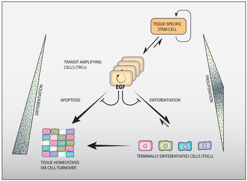
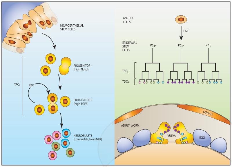
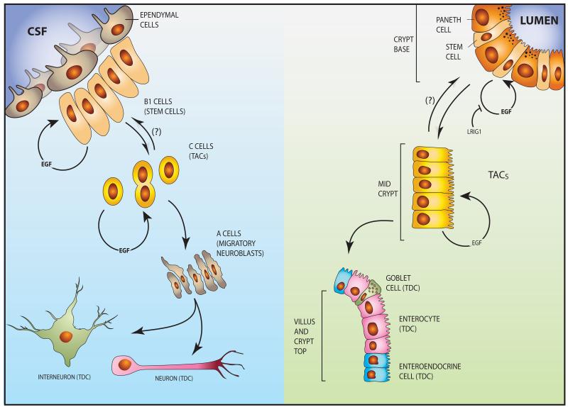
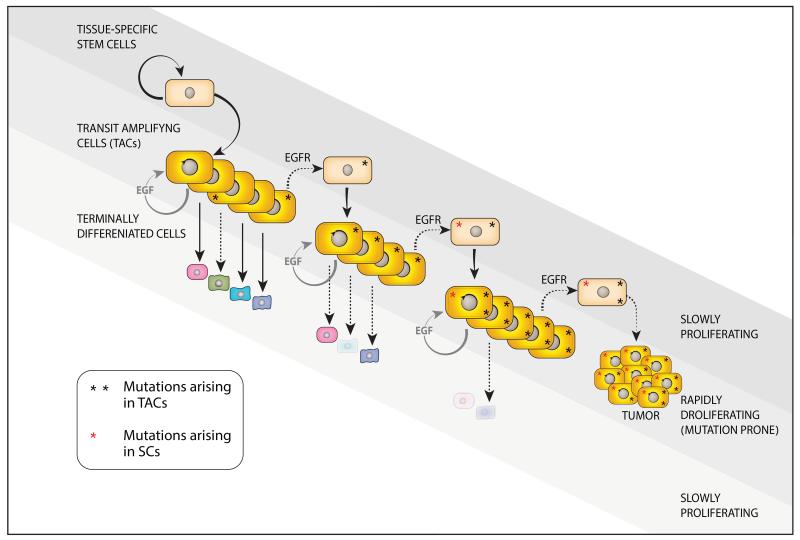
The epidermal growth factor receptor (EGFR) and a coreceptor denoted HER2/ERBB2 are frequently overexpressed or mutated in solid tumors, such as carcinomas and gliomas. In line with driver roles, cancer drugs intercepting EGFR or HER2 currently outnumber therapies targeting other hubs of signal transduction. To explain the roles for EGFR and HER2 as prime drivers and targets, we take lessons from invertebrates and refer to homeostatic regulation of several mammalian tissues. The model we infer ascribes to the EGFR-HER2 module pivotal functions in rapid clonal expansion of progenitors called transient amplifying cells (TACs). Accordingly, TACs of tumors suffer from replication stress, and hence accumulate mutations. In addition, several lines of evidence propose that in response to EGF and related mitogens, TACs might undergo dedifferentiation into tissue stem cells, which might enable entry of oncogenic mutations into the stem cell compartment. According to this view, antibodies or kinase inhibitors targeting EGFR-HER2 effectively retard some solid tumors because they arrest mutation-enriched TACs and possibly inhibit their dedifferentiation. Deeper understanding of the EGFR-HER2 module and relations between cancer stem cells and TACs will enhance our ability to control a broad spectrum of human malignancies.


Figures

Figure 1
Figure 1. The three-layered organization of cell turnover
Tissue-specific, adult stem cells are at the apex of the hierarchical three-layered organization. Their asymmetric divisions enable both self-renewal and generation of transit amplifying cells (TACs, or progenitors). TACs undergo several rounds of division (symbolized by a circular arrow), and then differentiate to form the various specialized cells of the tissue. Homeostatic regulation of tissues involves constant apoptosis and renewal, but stem cells are relatively resistant to apoptosis. Note that both stem cells and terminally differentiated cells (TDCs) are characterized by slow rates of mitoses, but the TACs are both short lived and rapidly proliferating. Expansion of the TAC compartment is governed, in some epithelial and neural organs, by the EGFR-HER2 module, which might inhibit both apoptosis and differentiation of TACs’ descendants. Importantly, the process is considered to be unidirectional. However, reversal (dedifferentiation) of TACs and TDC precursors might take place under certain conditions. For example, as discussed in this review, TACs might acquire some features of stemness once the EGFR-HER2 module is active.
Figure 2
Figure 2. Well-studied developmental processes of invertebrates are controlled by EGFR-driven progenitor cells
(Right panel) Vulval development in C. elegans is a multi-step process instigated by an inductive signal provided by the gonad-derived anchor cell (AC), in the form of LIN-3, the worm’s form of EGF. The secreted ligand travels a short distance to meet one of six equipotent stem cells, called vulva precursor cells (VPCs; normally P6.p), which express LET-23, the nematode EGFR. Following two divisions of the stimulated VPC and its two neighbors, which might be considered the vulval TACs, 22 differentiated cells are generated and form the vulva proper (lower panel). The other three VPCs form part of the body wall. (Left panel) In the Drosophila optic lobe, symmetrically dividing neuroepithelial stem cells transform into asymmetrically dividing neuroblasts, which later differentiate into many types of neurons. This sequential transformation involves two progenitor classes: type I progenitors are maintained by the Notch pathway, whereas type II progenitors are driven by active EGFRs. Transition to the neuroblast state and terminal differentiation is permitted by downregulation of the Notch and EGFR pathways,
Figure 3
Figure 3. Transit amplifying cells of the intestine and central nervous system of mammals are driven by EGFR-HER2 signals
(Right panel) The intestinal epithelium is organized into crypt and villus regions, with the stem cells and TACs localized to the crypt. Both quiescent and active stem cells exist in the base of the crypt next to Paneth cells, which provide essential niche signals, including specific EGFR ligands. EGFR is expressed by both SCs and TACs, and its mitogenic function toward SCs is strongly inhibited by LRIG1, a transmembrane molecule that physically binds with both EGFR and HER2. The TACs migrate toward the villus while undergoing EGFR-dependent mitoses followed by differentiation. The differentiation compartment, containing post-mitotic, lineage committed cells, such as goblet cells, enteroendocrine, tuft and Paneth cells, extends from the upper third of the crypt to the villus tip. Note that most differentiated cell populations migrate up the villi, but Paneth cells move downward. A reverse arrow and a question mark show putative dedifferentiation of TACs to SCs. (Left panel) A neural stem cell niche exists in the ventricular-subventricular zone (V-SVZ) in the lateral ventricles of mammalian brains. Ependymal cells line the ventricular surface and project cilia into the cerebrospinal fluid (CSF). The underlying type B1 cells are good candidates for the true adult neural stem cell identity. Type C cells are putative intermediate precursors (TACs) that differentiate to migrating neuroblasts (A cells). The latter divide and migrate out of the niche to form terminally differentiated neurons and interneurons. Note that both B1 (putative SCs) and C cells (TACs) are self-renewable and their mitoses are driven by EGFR signaling.
Figure 4
Figure 4. The EGFR-HER2 module might propel repeated cycles of TAC dedifferentiation to enable penetration of oncogenic mutations into the stem cell compartment and evolution of more aggressive tumors
The cellular hierarchy shown in Figure 1 is adopted here. The model presented assumes that active EGFR-HER2 of TACs expands these progenitors and, at the same time, promotes their dedifferentiation to stem cells. Due to their rapid rate of mitoses, the TACs, more than stem cells, suffer more from DNA damage generated endogenously by errors during DNA replication. Accordingly, the combination of frequent mutagenesis and dedifferentiation repeatedly feeds the stem cell compartment with more oncogenic mutations, which gives rise to progressively more aggressive tumors. Environmentally-induced and inborn mutations directly affecting adult SCs likely exacerbate tumor initiation and progression. Because the EGFR-HER2 module controls proliferation and probably also dediffentitaion of TACs, and these progenitors accumulate most mutations, it is predictable that pharmacological interceptors of the module would inhibit mutation accrual by SCs of epithelial and neural organs.

Similar articles

Cited by

References

    1. Pellettieri J, Sanchez Alvarado A. Cell turnover and adult tissue homeostasis: from humans to planarians. Annual review of genetics. 2007;41:83–105. - PubMed
    1. Tomasetti C, Vogelstein B. Cancer etiology. Variation in cancer risk among tissues can be explained by the number of stem cell divisions. Science. 2015;347:78–81. - PMC - PubMed
    1. Blanpain C, Horsley V, Fuchs E. Epithelial stem cells: turning over new leaves. Cell. 2007;128:445–458. - PMC - PubMed
    1. Hsu YC, Li L, Fuchs E. Emerging interactions between skin stem cells and their niches. Nature medicine. 2014;20:847–856. - PMC - PubMed
    1. Schneider MR, Wolf E. The epidermal growth factor receptor ligands at a glance. Journal of cellular physiology. 2009;218:460–466. - PubMed