Review
. 2015 Nov;36(11):782-797.
doi: 10.1016/j.tips.2015.07.004. Epub 2015 Oct 1.

Pharmacological Modulation of the N-End Rule Pathway and Its Therapeutic Implications

Affiliations
Review

Pharmacological Modulation of the N-End Rule Pathway and Its Therapeutic Implications

Jung Hoon Lee et al. Trends Pharmacol Sci. 2015 Nov.

Abstract

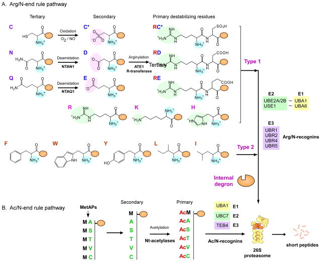
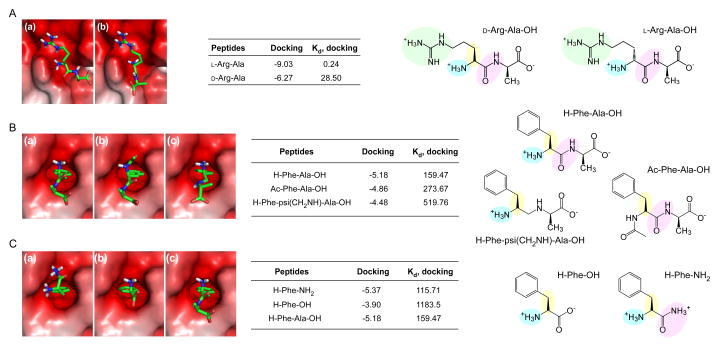
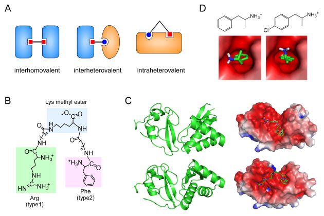
The N-end rule pathway is a proteolytic system in which single N-terminal amino acids of short-lived substrates determine their metabolic half-lives. Substrates of this pathway have been implicated in the pathogenesis of many diseases, including malignancies, neurodegeneration, and cardiovascular disorders. This review provides a comprehensive overview of current knowledge about the mechanism and functions of the N-end rule pathway. Pharmacological strategies for the modulation of target substrate degradation are also reviewed, with emphasis on their in vivo implications. Given the rapid advances in structural and biochemical understanding of the recognition components (N-recognins) of the N-end rule pathway, small-molecule inhibitors and activating ligands of N-recognins emerge as therapeutic agents with novel mechanisms of action.


Figures

Figure 1
Figure 1
Mammalian N-end rule pathway. (A) Arginylation branch of the N-end rule pathway (Arg/N-end rule pathway) in mammals. The tertiary destabilizing Cys residues are oxidized by O2 or nitric oxide (NO) into the secondary destabilizing residues Cys sulfinate (Cys-SO2) or Cys sulfonate (Cys-SO3). N-terminal Asn and Gln are deamidated into Asp and Glu by NTAN1 and NTAQ1, respectively. All of the secondary destabilizing residues expose negatively charged side chains (pink background). Secondary destabilizing residues, such as oxidized Cys, Asp, and Glu, are substrates of arginylation by ATE1 R-transferases. The positively charged side chains of N-terminal Arg, Lys, and His in type 1 destabilizing residues are shown in green. Type 2 destabilizing residues are hydrophobic residues such as Phe, Trp, Tyr, Leu, and Ile. These destabilizing residues are recognized and polyubiquitinated by Arg/N-recognins. In the Arg/N-end rule pathway, Ub can be activated and transferred by UBA1-UBE2A/2B (canonical) or UBA6-USE1 (noncanonical) cognates as E1–E2 systems. (B) Acetylation branch of the N-end rule pathway (Ac/N-end rule pathway) in mammals. N-terminal acetylation occurs at the newly exposed N-terminal residues, such as Ala, Ser, Thr, Val, and Cys, after the N-terminal Met excision by Met-endopeptidases (MetAPs) or at the retained N-terminal Met residue. These residues are recognized by the mammalian Ac/N-recognin TEB4.
Figure 2
Figure 2
Interaction between N-recognins and N-end rule pathway inhibitors (A) Binding modes calculated through in silico docking analysis of (a) L-Arg-Ala and (b) D-Arg-Ala with the UBR1 box (Protein Data Bank [PDB] code: 3NY3), and binding affinities (kcal/mol) and dissociation constants (μM) of the docked complexes. (B) Binding modes, binding affinities (kcal/mol), and dissociation constants (μM) of (a) Phe-Ala, (b) Ac-Phe-Ala, and (c) Phe-psi(CH2NH)-Ala with the ClpS domain (PDB code: 3DNJ). (C) Same as (B), except that Phe and its derivatives are used for the in silico docking analysis. Positively charged side chains, e.g., guanidino groups in Arg for interaction with the UBR box, are shown in green spheres in the chemical structures. Essential components of small molecules for interaction with UBR proteins, such as L-conformation, protonated α-amine groups, and amide bond characters, are shown in yellow, blue, and pink spheres, respectively.
Figure 3
Figure 3
Multivalent and monovalent inhibitors of the N-end rule pathway. (A) Types of multivalent ligands bound to their cognate proteins. (B) Structures of heterovalent molecule RF-Cn (where n indicates the length of hydrocarbon chains) tethering the core Lys methyl ester to the N-terminal Arg (type 1 destabilizing residue) and Phe (type 2), which are indicated by green and pink backgrounds, respectively. (C) Two possible structures of the UBR box-N-domain “combined” proteins (left) and their binding modes with the RF-C5 compound (right). Protein structures and docking models were obtained by using the Gramm-X protein-protein docking web server and semi-empirical PM6 method with the Gaussian 09 program [30]. (D) Chemical structures of amphetamine (left) and para-chloroamphetamine (right), and their binding modes with the N-domain (PDB code: 3DNJ) calculated with a ligand-receptor docking computation by using AutoDock version 4.2 with the Lamarckian genetic algorithm [85].

References

    1. Choi AM, et al. Autophagy in human health and disease. N Engl J Med. 2013;368:651–662. - PubMed
    1. Schwartz AL, Ciechanover A. The ubiquitin-proteasome pathway and pathogenesis of human diseases. Annu Rev Med. 1999;50:57–74. - PubMed
    1. Lee MJ, et al. Tau degradation: the ubiquitin-proteasome system versus the autophagy-lysosome system. Prog Neurobiol. 2013;105:49–59. - PubMed
    1. Glickman MH, Ciechanover A. The ubiquitin-proteasome proteolytic pathway: destruction for the sake of construction. Physiol Rev. 2002;82:373–428. - PubMed
    1. Kamura T, et al. Rbx1, a component of the VHL tumor suppressor complex and SCF ubiquitin ligase. Science. 1999;284:657–661. - PubMed

Publication types

LinkOut - more resources