. 2015 Oct 5:5:14733.
doi: 10.1038/srep14733.

USP15 regulates SMURF2 kinetics through C-lobe mediated deubiquitination

Affiliations

USP15 regulates SMURF2 kinetics through C-lobe mediated deubiquitination

Prasanna Vasudevan Iyengar et al. Sci Rep. .

Abstract

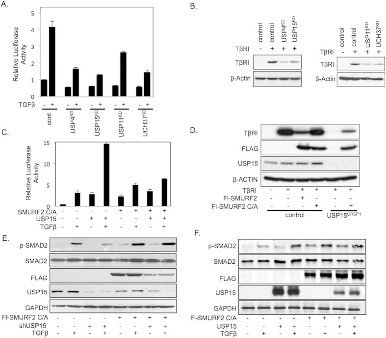
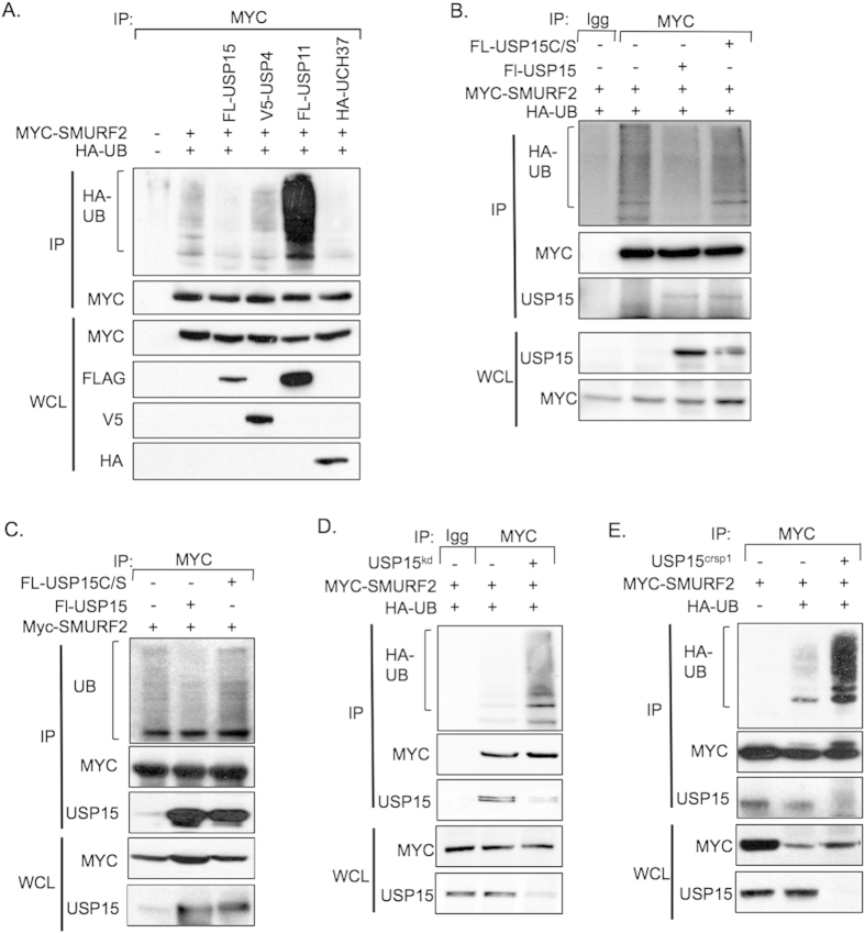
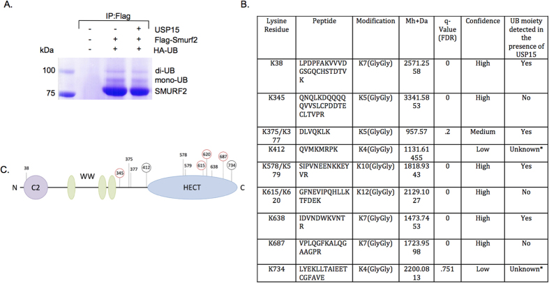
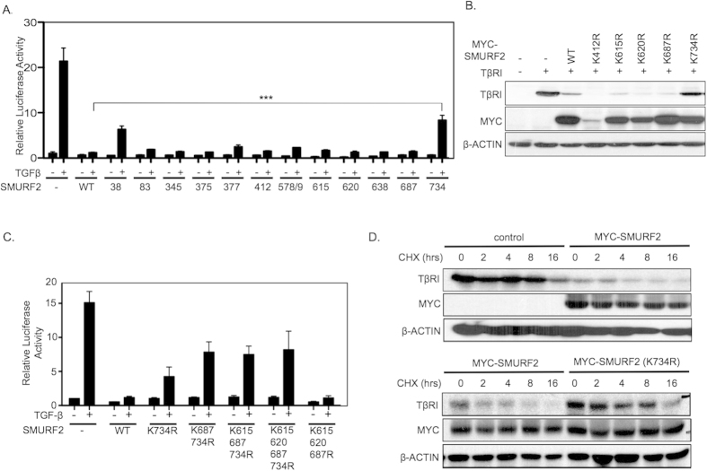
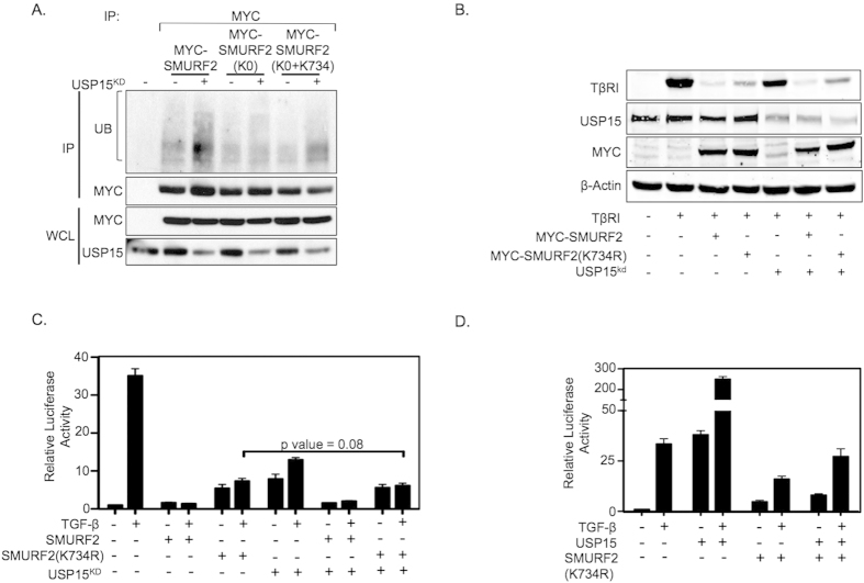
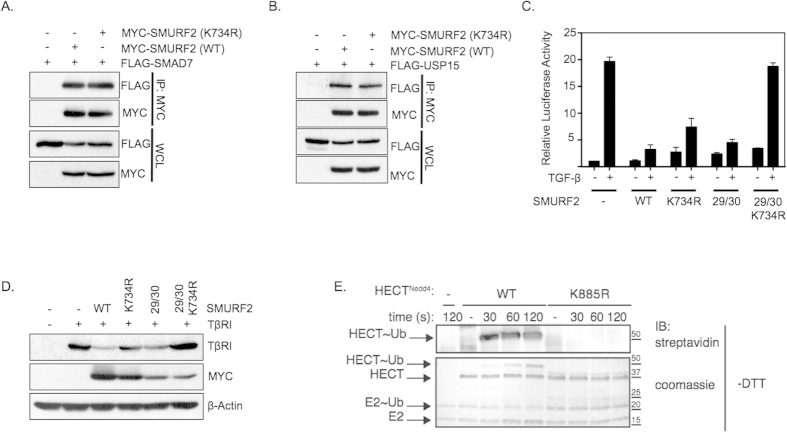
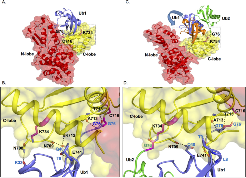
Ubiquitin modification of the TGF-β pathway components is emerging as a key mechanism of TGF-β pathway regulation. To limit TGF-β responses, TGF-β signaling is regulated through a negative feedback loop whereby the E3 ligase SMURF2 targets the TGF-β receptor (TβR) complex for ubiquitin-mediated degradation. Counteracting this process, a number of deubiquitinating (DUBs) enzymes have recently been identified that deubiquitinate and stabilize the TβR. However the precise mechanism by which these DUBs act on TβR function remains poorly defined. Here, we demonstrate that apart from targeting the TβR complex directly, USP15 also deubiquitinates SMURF2 resulting in enhanced TβR stability and downstream pathway activation. Through proteomic analysis, we show that USP15 modulates the ubiquitination of Lys734, a residue required for SMURF2 catalytic activity. Our results show that SMURF2 is a critical target of USP15 in the TGF-β pathway and may also explain how USP15 and SMURF2 target multiple complementary protein complexes in other pathways.


Figures

Figure 1
Figure 1. USP15 function in the TGF-β pathway is dependent on SMURF2 in HEK293T cells.
(A) Luciferase assay in HEK293T cells transfected with CAGA-luciferase reporter and shRNAs targeting deubiquitinating enzymes USP4, USP11, USP15, or UCH37 treated with TGF-β (2.5 ng/ml). Data are mean ± s.d. (B) Immunoblot analysis showing levels of transfected TβRI along with shRNA vectors described in A. (C) Luciferase assay representing TGF-β activity in 293T cells transfected with CAGA-luciferase reporter and either catalytically inactive mutant of SMURF2 C/A, USP15, or both treated with TGF-β (2.5 ng/ml). Data are mean ± s.d. (D) Immunoblot analysis in 293T or USP15CRSP1 cells expressing TβRI and Flag-tagged SMURF2 or SMURF2 C/A. Whole cell lysates were probed with the indicated antibodies. (E) Immunoblot analysis with HEK293T cells showing changes in p-SMAD2 levels when transfected with shRNA targeting USP15, SMURF2 C/A or both and treated with TGF-β overnight (2.5 ng/ml). (F) Immunoblot analysis showing changes in p-SMAD2 levels when transfected with USP15, SMURF2 (C/A) or both and treated with TGF-β overnight (2.5 ng/ml).
Figure 2
Figure 2. USP15 targets SMURF2 for deubiquitination.
(A) Immunoprecipitation with anti-Myc antibodies in HEK293T cells overexpressing Myc-SMURF2, the indicated deubiquitinating enzymes and hemagglutinin (HA)-tagged ubiquitin (HA-UB). Immunoblot analysis probed for the indicated proteins is shown. (B) Immunoprecipitation analysis with 293T cells expressing SMURF2, USP15 or USP15 C/S and HA-UB; an immunoblot analysis of the indicated proteins is shown. (C) Immunoprecipitation analysis with 293T cells expressing SMURF2 and USP15 or USP15 C/S; an immunoblot analysis of the indicated proteins is shown. (D) Immunoprecipitation in HEK293T transfected with Myc-SMURF2, HA-ubiquitin and an shRNA against USP15, cells were immunoprecipitated with either mouse IgG or Myc antibodies; immunoblot analysis of the indicated proteins is shown. (E) Immunoprecipitation with anti-Myc antibodies using control or USP15CRISPR cell lines, overexpressing Myc-SMURF2 and HA-ubiquitin.
Figure 3
Figure 3. USP15 deubiquitinates multiple lysine residues on SMURF2.
(A) Immunoprecipitation using anti-Flag resin in HEK293T cells, transfected with control vector (lane 1) or Flag-SMURF2 (lanes 2 and 3), K0-ubiquitin, and SMAD7 with or without USP15 overexpression and visualized using coomassie staining; bands above Flag-SMURF2 show mono and di-mono-ubiquitinated Flag-SMURF2. (B) Table showing candidate ubiquitinated lysine residues derived from mass spectrometric analysis. *Indicates non-detectable peptides by MASS SPEC. (C) Schematic representation of SMURF2 with its domains and candidate ubiquitination sites; ubiquitinated sites not detected (red circle) or detected with low confidence (black circle) in the presence of USP15 are indicated.
Figure 4
Figure 4. Lysine 734 of SMURF2 is critical for SMURF2 function.
(A) Luciferase assay showing relative TGF-β activity in HEK293T cells transfected with CAGA-Luc and either wild-type SMURF2 or SMURF2 mutants representing candidate ubiquitin sites and treated with TGF-β overnight (2.5 ng/ml). ***P value = 0.0002 using Student’s t test. Data are mean ± s.d. (B) Immunoblot analysis in HEK293T cells expressing TβRI in the presence of either wild-type SMURF2 or SMURF2 mutants as described in A. (C) Luciferase assay in HEK293T cells transfected with CAGA-luciferase reporter with wild-type SMURF2, SMURF2 K734R or in combination with other candidate deubiquitination sites of the HECT domain of SMURF2 and treated with TGF-β overnight (2.5 ng/ml). Data are mean ± s.d. (D) HEK293T cells were transfected with TβRI and either control, Myc-SMURF2 or Myc-SMURF2 K734R; the cells were treated with cycloheximide (10 μg/ml) and TGF-β (2.5 ng/ml) and lysed at the indicated time points.
Figure 5
Figure 5. USP15 exerts its effect on SMURF2 through lysine 734.
(A) Immunoprecipitation analysis with anti-Myc antibodies in HEK293T cells expressing either wild-type Myc-SMURF2 or Myc-SMURF2 with all the candidate deubiquitination sites mutated including or excluding lysine 734, indicated by K0 or K0-K734, respectively. (B) Immunoblot analysis using HEK293T cells transfected with the indicated plasmids, β-actin is shown as loading control. (C) Luciferase assay representing TGF-β activity in HEK293T cells transfected with the indicated plasmids either alone or together with knockdown vectors targeting USP15 and treated with TGF-β overnight (2.5 ng/ml). P value = 0.08 using Student’s t test. Data are mean ± s.d. (D) Luciferase assay representing TGF-β activity in HEK293T cells transfected with the indicated plasmids either alone or together with ectopic expression of USP15 and treated with TGF-β overnight (2.5 ng/ml). Data are mean ± s.d.
Figure 6
Figure 6. Lysine 734 of SMURF2 is required for SMURF2 transthiolation.
(A) Immunoprecipitation with anti-Myc antibodies in HEK293T cells transfected with Flag-SMAD7 and either with Myc-SMURF2 or Myc-SMURF2 (K734R); whole cell lysates are indicated below (WCL). (B) Immunoprecipitation with anti-Myc antibodies in HEK293T cells transfected with Flag-USP15 and either with Myc-SMURF2 or Myc-SMURF2 (K734R). (C) Luciferase activity in HEK293T cells transfected with CAGA-luciferase, Myc-SMURF2, Myc-SMURF2 (K734R), Myc-SMURF2 (FF29/30AA) or Myc-SMURF2 (FF29/30AA+K734R) treated with TGF-β (2.5 ng/ml). Data are mean ± s.d. (D) Aforementioned plasmids were co-transfected with TβRI in 293T cells; an immunoblot analysis of the indicated proteins is shown. (E) An in vitro trans-thiolation assay; purified HECT domain of NEDD4 (either wild-type or K885R) was incubated with E1, E2 and ubiquitin for the indicated time points and analyzed through immunoblotting or coomassie staining.
Figure 7
Figure 7. Mutating lysine 734 of SMURF2 mitigates TGF-β induced migration.
(A) MDA-MB-231 cells stably expressing SMURF2 (wild-type) or SMURF2 (K734R) were plated for a scratch assay and treated with SB431542 (5 μM) or TGF-β (5 ng/ml), panels show migration at 0 and 24 hours. (B) Percentage of migrated area within the black dotted lines was estimated with respect to control (0 hour) and a graph was plotted. *P value = 0.014 using Student’s t test. Data are mean ± s.d. (C) Immunoblotting showing lentiviral induced SMURF2 (wild-type) or SMURF2 (K734R) expression in MDA-MB-231 cells. (D) Transwell assay of MDA-MB-231 cells infected with SMURF2 or SMURF2 (K734R) and treated with SB431542 (5 μM) or TGF-β (5 ng/ml) for 16 hours before fixation and staining with crystal violet. (E) Graph represents total number of migrated cells from four random fields from Figure D. *P value = 0.011 using Student’s t test. Data are mean ± s.d.
Figure 8
Figure 8. Model for Ub-loaded SMURF2 HECT domain in complex with Ub bound at lysine 734.
(A) Molecular model of donor ubiquitin (Ub1, blue) bound to the catalytic pocket of HECT domain of SMURF2. (B) Inset of A showing the details of the hydrogen-bond interaction between SMURF2 C-Lobe (yellow) and catalytically bound Ub1 (blue). Thioester bond between Gly76 and Cys716 is denoted. Unbound Lys 734 is also shown. (C) Molecular model of ubiquitin (green, Ub2) bound to Lys734 site with donor ubiquitin (Ub1) bound to the catalytic pocket. The displacement of Ub1 (shown in blue and orange) due to the presence of Ub2 is indicated by an arrow. (D) Inset of C showing the hydrogen-bond interactions between SMURF2 C-Lobe (yellow) and Ub1 (blue) in the presence of Lys734 bound Ub2 (green). The isopeptide bond between Lys734 and Gly76 of Ub2 is also shown.

References

    1. Deshaies R. J. & Joazeiro C. A. RING domain E3 ubiquitin ligases. Annual review of biochemistry 78, 399–434, 10.1146/annurev.biochem.78.101807.093809 (2009). - DOI - PubMed
    1. Scheffner M., Nuber U. & Huibregtse J. M. Protein ubiquitination involving an E1-E2-E3 enzyme ubiquitin thioester cascade. Nature 373, 81–83, doi: 10.1038/373081a0 (1995). - PubMed
    1. Massague J. TGFbeta in Cancer. Cell 134, 215–230, 10.1016/j.cell.2008.07.001 (2008). - DOI - PMC - PubMed
    1. Massague J., Seoane J. & Wotton D. Smad transcription factors. Genes & development 19, 2783–2810, 10.1101/gad.1350705 (2005). - DOI - PubMed
    1. Kavsak P. et al. Smad7 binds to Smurf2 to form an E3 ubiquitin ligase that targets the TGF beta receptor for degradation. Molecular cell 6, 1365–1375 (2000). - PubMed

Publication types

LinkOut - more resources