Review
. 2015;6(2):10.1128/ecosalplus.ESP-0010-2014.
doi: 10.1128/ecosalplus.ESP-0010-2014.

DNA Topoisomerases

Review

DNA Topoisomerases

Natassja G Bush et al. EcoSal Plus. 2015.

Abstract

DNA topoisomerases are enzymes that control the topology of DNA in all cells. There are two types, I and II, classified according to whether they make transient single- or double-stranded breaks in DNA. Their reactions generally involve the passage of a single- or double-strand segment of DNA through this transient break, stabilized by DNA-protein covalent bonds. All topoisomerases can relax DNA, but DNA gyrase, present in all bacteria, can also introduce supercoils into DNA. Because of their essentiality in all cells and the fact that their reactions proceed via DNA breaks, topoisomerases have become important drug targets; the bacterial enzymes are key targets for antibacterial agents. This article discusses the structure and mechanism of topoisomerases and their roles in the bacterial cell. Targeting of the bacterial topoisomerases by inhibitors, including antibiotics in clinical use, is also discussed.


Figures
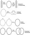
Figure 1
Figure 1
Reactions performed by type I topoisomerases. Examples of specific type I topoisomerases that catalyze the indicated reactions are given above the arrows. It is important to note that in the decatenation/catenation reaction, the non-nicked plasmid may be supercoiled before decatenation/catenation occurs; for illustrative purposes it has been drawn as relaxed. (Adapted from reference with permission of the publisher.) doi:10.1128/ecosalplus.ESP-0010-2014.f1
Figure 2
Figure 2
Reactions performed by type II topoisomerases. Examples of specific type II topoisomerases that catalyze the indicated reactions are given above the arrows. It is important to note that in the decatenation/catenation reaction, the plasmids may be supercoiled before decatenation/catenation occurs; for illustrative purposes they have been drawn as relaxed. Although only relaxation of negative supercoils is shown, all known type II topoisomerases can relax positively supercoiled DNA as well. (Redrawn from reference with permission of the publisher.) doi:10.1128/ecosalplus.ESP-0010-2014.f2
Figure 3
Figure 3
Model of the topology of a replicating chromosome. The chromosome is separated into domains with the boundaries represented as orange boxes; the replication fork is in the center. Positive supercoiling occurs ahead of the replication fork, and precatenanes may form behind it. (Reprinted from reference . Copyright 2001 National Academy of Sciences, U.S.A.) doi:10.1128/ecosalplus.ESP-0010-2014.f3
Figure 4
Figure 4
Formation of catenated DNA at the termination of replication. (a and b) Converging replication forks (a) lead to the interwinding of daughter molecules and the formation of precatenanes (b). (c) Upon the completion of replication, the products are catenated DNA circles. (Reprinted from reference with permission of the publisher.) doi:10.1128/ecosalplus.ESP-0010-2014.f4
Figure 5
Figure 5
Primary domain structures of type I topoisomerases. Black bars indicate catalytic residues. Y is the catalytic tyrosine which forms the covalent bond with the phosphodiester backbone of the cleaved single-strand of DNA (319 in E. coli topo I, 328 in E. coli topo III, 809 in A. fulgidus reverse gyrase, 723 in human topo I, and 226 in M. kandleri topo V) (for a full description of all catalytic residues, see reference 147). In type IB, NTD is the N-terminal domain, CTD is the C-terminal domain. In type IC, HTH is helix-turn-helix, HhH is helix-hairpin-helix. (Adapted from reference . Schoeffler AJ, Berger JM. 2008. DNA topoisomerases: harnessing and constraining energy to govern chromosome topology. Q Rev Biophys 41:41–101. © Cambridge University Press, reproduced with permission.) doi:10.1128/ecosalplus.ESP-0010-2014.f5
Figure 6
Figure 6
Primary domain structures of type II topoisomerases. Black bars indicate catalytic residues. Y is the catalytic tyrosine which forms the covalent bond with the phosphodiester backbone of the cleaved strand of DNA (782 in S. cerevisiae topo II, 122 in E. coli DNA gyrase, 120 in E. coli topo IV, and 105 in M. mazei topo VI) (for full description of all catalytic residues, see reference 148). GHKL is the ATPase domain, TOPRIM stands for topoisomerase/primase domain, WHD is the winged-helix domain, CTD is the C-terminal domain, H2tH is the helix-helix-turn helix domain, and Ig is an immunoglobulin-type fold (not seen in all species). (Adapted from reference . Schoeffler AJ, Berger JM. 2008. DNA topoisomerases: harnessing and constraining energy to govern chromosome topology. Q Rev Biophys 41:41–101. © Cambridge University Press, reproduced with permission.) doi:10.1128/ecosalplus.ESP-0010-2014.f6
Figure 7
Figure 7
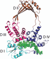
Structure of an N-terminal fragment of E. coli DNA topoisomerase I in a covalent complex with DNA. A ribbon representation of the overall structure of the protein is presented, with four subdomains (DI to DIV) shown in different colors. The bound DNA is shown in green as an electron density map. (Reprinted from reference with permission of the publisher.) doi:10.1128/ecosalplus.ESP-0010-2014.f7
Figure 8
Figure 8
Proposed mechanism for E. coli topo I. The enzyme binds DNA (T segment in red, G segment in black; not to scale) and cleaves one strand (active-site tyrosine in purple), forming a 5′-phosphodiester linkage. The complementary strand is passed through the gap and into the central cavity of the enzyme. The light blue circles indicate areas of structural changes during the open conformation of the enzyme. The nick is resealed, and the strand is released. It is possible that the cycle proceeds in reverse with the T segment being passed out of the enzyme rather than in (steps 7 through 1 rather than 1 through 7) (24). (This figure was published in Viard T, de la Tour CB. 2007. Type IA topoisomerases: a simple puzzle? Biochimie 89:456–467. Copyright © 2007 Elsevier Masson SAS. [314] All rights reserved.) doi:10.1128/ecosalplus.ESP-0010-2014.f8
Figure 9
Figure 9
Structure of truncated (amino acids 1 through 1177) S. cerevisiae topo II bound to DNA and ADPNP. One monomer is shaded grey, and the other is colored by functional region. WHD is the winged-helix domain, TOPRIM is the topoisomerase-primase domain. The black box indicates the position of ADPNP, and green indicates DNA. (Reprinted from reference with permission of the publisher.) doi:10.1128/ecosalplus.ESP-0010-2014.f9
Figure 10
Figure 10
Structures of topoisomerase IV. (A) Structure of the ParE-ParC55 fusion construct (122) (PDB: 4I3H). Yellow indicates the GHKL domain, orange is the transducer domain, teal is the winged-helix domain (WHD), purple is the tower domain, and blue shows the coiled-coil domain (see Fig. 6 for domain structure). (B) Space-filled model of the structure shown in panel A. (C) ParE 43-kDa N-terminal fragment complexed with ADPNP (black box) (PDB: 1S16) (114). It is proposed that the open conformation of ParE as seen in panel A is the conformation pre-ATP binding whereas the conformation seen in panel C is the post-ATP-binding conformation. (D) ParC C-terminal domain in two orientations (PDB: 1ZVT) (115). doi:10.1128/ecosalplus.ESP-0010-2014.f10
Figure 11
Figure 11
Structures of DNA gyrase. (A) Model of the full-length structure of DNA gyrase. Yellow indicates the GHKL domain, orange is the transducer domain, teal is the winged-helix domain (WHD), purple is the tower domain, blue shows the coiled-coil domain, and pink indicates the C-terminal domain (see Fig. 6 for the domain structure). The full-length protein structure was modeled on the GyrB 43-kDa fragment (PDB: 1EI1), a B-A fusion construct (PDB: 3NUH) (144), and the GyrA 35-kDa C-terminal domain (PDB: 3L6V). (B) Space-filled model of the structure shown in panel A. (C) Four principal domains of gyrase. 1 is the E. coli GyrB 43-kDa fragment complexed with ADPNP; 2 is the E. coli GyrB TOPRIM domain; 3 is the E. coli GyrA 59-kDa subunit; 4 is the E. coli GyrA C-terminal domain in two orientations (PDB: 1ZI0). doi:10.1128/ecosalplus.ESP-0010-2014.f11
Figure 12
Figure 12
Cryo-EM map of the DNA-bound complex modeled with the crystal structures of DNA gyrase domains alone (A) and with duplex DNA (B). In particular, the crystal structures of the ATPase (PDB:1EI1) and the DNA-binding-cleavage domain in the presence of ciprofloxacin (PDB:2XCT) were modeled into the core of the map with the two additional densities on both side of the core enzyme accommodating the C-terminal domains (PDB:3L6V). (Reprinted from reference with permission of the publisher.) doi:10.1128/ecosalplus.ESP-0010-2014.f12
Figure 13
Figure 13
Model for negative supercoiling by DNA gyrase. The domains are colored as follows: GyrB43, dark blue; GyrB TOPRIM, red; GyrB tail, green; GyrA59, orange; GyrA C-terminal domain, light blue. The G and T DNA segments are colored black and purple, respectively. 1, subunits and DNA in their proposed free states in solution. Stars indicate the active-site residues for DNA cleavage, and the circle indicates the ATP-binding pocket. 2, The G segment binds across GyrA at the dimer interface, and the GyrA C-terminal domain wraps the DNA to present the T segment in a positive crossover. 3, ATP is bound, which closes the GyrB clamp capturing the T segment, and the G segment is transiently cleaved. 4, Hydrolysis of one ATP molecule allows GyrB to rotate, the DNA gate to widen, and the T segment to be transported through the cleaved G segment. 5, The T segment exits through the C gate, and the G segment is religated. The hydrolysis of the remaining ATP molecule resets the enzyme. The right panel shows the side view for illustrations 2 through 4. (Reprinted from reference with permission of the publisher.) doi:10.1128/ecosalplus.ESP-0010-2014.f13
Figure 14
Figure 14
Structures of aminocoumarins. doi:10.1128/ecosalplus.ESP-0010-2014.f14
Figure 15
Figure 15
The binding sites of novobiocin and ADPNP in GyrB partially overlap. Part of the N-terminal GyrB structure is shown, with ADPNP in red and novobiocin in blue (204). doi:10.1128/ecosalplus.ESP-0010-2014.f15
Figure 16
Figure 16
Structure of simocyclinone D8. doi:10.1128/ecosalplus.ESP-0010-2014.f16
Figure 17a
Figure 17a
Structure of the N-terminal domain of GyrA (GyrA55) complexed with simocyclinone D8. The protein dimer is shown in gold and blue (ribbon representation), and the bound simocyclinone D8 dimer is shown in space-filling representation. (A) Side view. (B) Top view. Note that the polyketide end of each simocyclinone molecule also binds to the other monomer across the dimer (DNA-gate) interface (221). doi:10.1128/ecosalplus.ESP-0010-2014.f17a
Figure 18
Figure 18
Chemical structures of a selection of quinolones. Quinolones are divided into generations based on their antibacterial spectrum. The first-generation drugs (e.g., nalidixic acid and oxolinic acid) are examples of older acidic (narrow-spectrum) quinolones, whereas the higher-generation drugs (e.g., ciprofloxacin, sparfloxacin, and gatifloxacin) are examples of the amphoteric fluoroquinolones (expanded-spectrum compounds) (315, 316). doi:10.1128/ecosalplus.ESP-0010-2014.f18
Figure 19
Figure 19
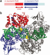
Topo IV-DNA-quinolone complex. (a) ParE28-ParC58 is a fusion of the C-terminal region of ParE and the N-terminal region of ParC. (b) ParE28-ParC58 complex with DNA (green), moxifloxacin (yellow carbons), and Mg2+ ions (orange spheres). One fused ParE-ParC subunit is shown in red-blue; the other is all gray. (Reprinted from reference with permission of the publisher.) doi:10.1128/ecosalplus.ESP-0010-2014.f19
Figure 20
Figure 20
Structure of the DNA gyrase inhibitor MccB17. doi:10.1128/ecosalplus.ESP-0010-2014.f20
Figure 21
Figure 21
Structure of CcdB and its complex with GyrA. (A) Crystal structure of GyrA59 with and without CcdB. The left panel shows the apo-structure. The green spheres indicate the positions of residues that, if mutated, confer resistance to CcdB (for an overview of these mutations, see reference 285). The right panel shows GyrA59 bound to CcdB (GyrA59 dimer colored blue and orange by subunit; CcdB dimer colored green and purple). (B) Diagram of the proposed mode of action of CcdB. The GyrB subunits are shown in yellow, the GyrA subunits are shown in green, and CcdB is shown in pink. The T segment is shown in red, and the G segment is shown in black; * represents ATP. After DNA binding and opening of the DNA gate, CcdB binds to the GyrA, blocking the T segment from exiting the C gate, and stabilizes the cleavage complex by stalling the enzyme in the open conformation. In this state there may be a futile ATP hydrolysis event which would fail to reset the enzyme. While only one ATP molecule is shown in the model, it is possible that at this stage two molecules of ATP may be bound (for the full catalytic cycle, see Fig. 13). Although this figure shows ATP binding during inhibition by CcdB, it has been shown that CcdB can stabilize the cleavage complex in the absence of ATP (285). (Redrawn from reference with permission from the publisher.) doi:10.1128/ecosalplus.ESP-0010-2014.f21
Figure 17b
Figure 17b
Structure of the N-terminal domain of GyrA (GyrA55) complexed with simocyclinone D8. The protein dimer is shown in gold and blue (ribbon representation), and the bound simocyclinone D8 dimer is shown in space-filling representation. (A) Side view. (B) Top view. Note that the polyketide end of each simocyclinone molecule also binds to the other monomer across the dimer (DNA-gate) interface (221). doi:10.1128/ecosalplus.ESP-0010-2014.f17b

References

    1. Watson JD, Crick FH. 1953. Genetical implications of the structure of deoxyribonucleic acid. Nature 171:964–967. [PubMed]10.1038/171964b0 - DOI - PubMed
    1. Bates AD, Maxwell A. 2005. DNA Topology. Oxford University Press, Oxford, United Kingdom.
    1. Deweese JE, Osheroff MA, Osheroff N. 2008. DNA topology and topoisomerases: teaching a “knotty” subject. Biochem Mol Biol Educ 37:2–10. [PubMed]10.1002/bmb.20244 - DOI - PMC - PubMed
    1. Sissi C, Palumbo M. 2010. In front of and behind the replication fork: bacterial type IIA topoisomerases. Cell Mol Life Sci 67:2001–2024. [PubMed]10.1007/s00018-010-0299-5 - DOI - PMC - PubMed
    1. Vos SM, Tretter EM, Schmidt BH, Berger JM. 2011. All tangled up: how cells direct, manage and exploit topoisomerase function. Nat Rev Mol Cell Biol 12:827–841. [PubMed]10.1038/nrm3228 - DOI - PMC - PubMed

Publication types

MeSH terms

LinkOut - more resources