

OXIDATIVE STRESS IN HUMAN LEUKEMIA (HL-60), HUMAN LIVER CARCINOMA (HepG2), AND HUMAN (JURKAT-T) CELLS EXPOSED TO ARSENIC TRIOXIDE
- PMID: 26435679
- PMCID: PMC4589149
OXIDATIVE STRESS IN HUMAN LEUKEMIA (HL-60), HUMAN LIVER CARCINOMA (HepG2), AND HUMAN (JURKAT-T) CELLS EXPOSED TO ARSENIC TRIOXIDE
Abstract
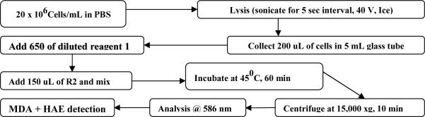
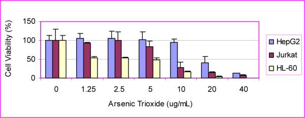
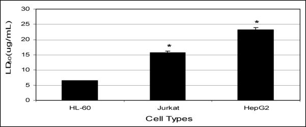
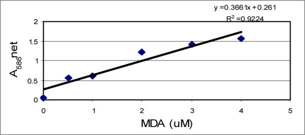
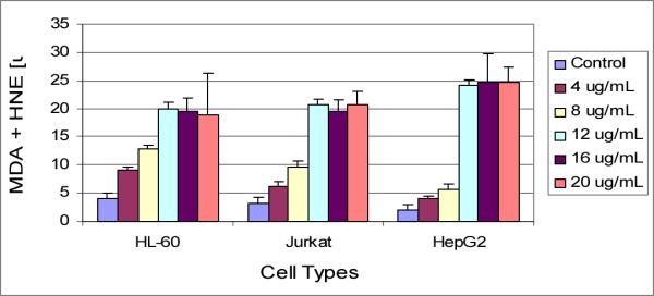
Recent studies have shown that arsenic trioxide can induce a clinical remission in patients with acute promyelocytic leukemia. However, the molecular mechanisms of action remain to be elucidated. In this research, we performed the MTT assay to evaluate the cytotoxic effects of arsenic trioxide (ATO) to HL-60 cells and to compare their relative sensitivity to that of HepG2, and Jurkat T cells. We also performed the thiobarbituric acid test to determine the levels of malondialdehyde (MDA) plus 4-hydroxy-2 (E)-nonenal (4-HAE) production in these three cell lines following exposure to arsenic trioxide. The result of MTT assay clearly demonstrated that ATO has a significant cytotoxic effect on HL-60, Jurkat, and HepG2 cells; showing 24 hrs LD50 values of 6.4 ± 0.6 μg/mL, 15 ± 3.84 μg/mL, and 23.2 ± 6.03 μg/mL, respectively. These data indicated that HL-60 cells are about twice as sensitive to arsenic toxicity compared to Jurkat T cells and about 3 times more sensitive to arsenic trioxide compared to HepG2 cells. The result of the thiobarbituric acid test demonstrated that arsenic trioxide treatment resulted in a significant increase (p <0.05) of MDA and HAE production, indicating that oxidative stress plays a key role in arsenic induced toxicity and cell injury. MDA and HAE levels were significantly higher in arsenic trioxide-treated HL-60 cells, indicating that these cells appear to be more sensitive to oxidative stress than HepG2 and Jurkat T- cells. In summary, these results indicate that the pharmacology of ATO as an effective anti-cancer drug is associated with its cytotoxic effects in human promyelocytic leukemic cells. This cytotoxicity is found to be mediated by oxidative stress, a biomarker of cellular injury.
Keywords: Arsenic trioxide; HAE; HL-60; HepG2; Jurkat; MDA; lipid peroxidation.
Figures
References
-
- Zhang K, Ohnishi K, Shigeno K, Fujisawa S, Naito K, Nakamura S, Takeshita K, Takeshita A, Ohno R. The induction of apoptosis and cell cycle arrest by arsenic trioxide in lymphoid neoplasms. Leukemia (Baltimore) 1998;12:1383–1391. - PubMed
-
- Rousselot P, Labaume S, Marolleau JP, Larghero J, Noguera MK, Brouet JC, Fermand JP. Arsenic trioxide and melarsoprol induce apoptosis in plasma cell lines and in plasma cells from myeloma patients. Cancer Res. 1999;59:1041–1048. - PubMed
-
- Li JH, Rossman TC. Inhibition of DNA ligase activity by arsenite: A possible mechanism of its comutagenesis. Mol. Toxicol. 1989;2:1–9. - PubMed
-
- Chen C-S, Siegel DM. Arsenical keratosis. eMedicine J. 2001;2(6)
-
- Jai P, Chen G, Huang X, Cai X, Yang J, Wang L, Zhou Y, Shen Y, Zhou L, Yu Y, Chen S, Zhang X, Wang Z. Arsenic trioxide induces multiple myeloma cell apoptosis via disruption of mitochondrial transmembrane potentials and activation of caspace-3. Chin. Med. J. (Engl) 1999;(114):19–24. - PubMed
Grants and funding
LinkOut - more resources
Full Text Sources
Research Materials