Review
. 2015:2015:816856.
doi: 10.1155/2015/816856. Epub 2015 Sep 8.

Review of the Structural and Dynamic Mechanisms of PPARγ Partial Agonism

Affiliations
Review

Review of the Structural and Dynamic Mechanisms of PPARγ Partial Agonism

Alice J Kroker et al. PPAR Res. 2015.

Abstract

PPARγ (peroxisome proliferator activated receptor γ) is a ligand activated transcription factor of the nuclear receptor superfamily that controls the expression of a variety of genes involved in fatty acid metabolism, adipogenesis, and insulin sensitivity. While endogenous ligands of PPARγ include fatty acids and eicosanoids, synthetic full agonists of the receptor, including members of the thiazolidinedione (TZD) class, have been widely prescribed for the treatment of type II diabetes mellitus (T2DM). Unfortunately, the use of full agonists has been hampered by harsh side effects with some removed from the market in many countries. In contrast, partial agonists of PPARγ have been shown to retain favourable insulin sensitizing effects while exhibiting little to no side effects and thus represent a new potential class of therapeutics for the treatment of T2DM. Partial agonists have been found to not only display differences in transcriptional and cellular outcomes, but also act through distinct structural and dynamic mechanisms within the ligand binding cavity compared to full agonists.


Figures

Figure 1
Figure 1
PPARγ domain organization. (a) Primary structure of PPARγ. (b) Crystal structure of the intact PPARγ-RXR heterodimer bound to PPRE DNA with agonist ligands retinoic acid and full agonist rosiglitazone. Proteins and DNA are shown as ribbons while ligands are shown as spheres. RXR is coloured red, the PPARγ ligand binding domain is coloured yellow, the PPARγ DNA binding domain is coloured green, the PPARγ hinge is coloured cyan, and the NCOA2 coactivator peptide is coloured blue. PDB: 3DZY [14].
Figure 2
Figure 2
Interaction mode of full agonist rosiglitazone. The PPARγ ligand binding domain is shown in yellow and rosiglitazone is shown in white sticks coloured by element. (a) Ribbons diagram of the PPARγ ligand binding domain bound to rosiglitazone. The branches of the ligand binding pocket have been labelled with Roman numerals. (b) Hydrogen bond network of the rosiglitazone TZD head group with AF2 residues. PDB: 4EMA [49].
Figure 3
Figure 3
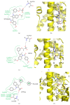
Crystal structures of indole containing PPARγ partial agonists. (a) Poseview map of SPPARγM2. (b) Crystal structure of SPPARγM2 bound to the PPARγ ligand binding domain. PDB: 2P4Y [50]. (c) Poseview map of MRL24. (d) Crystal structure of MRL24 bound to the PPARγ ligand binding domain. PDB: 2Q5P [47]. (e) Poseview map of nTZDpa. (f) Crystal structure of nTZDpa bound to the PPARγ ligand binding domain. PDB: 2Q5S [47]. The branches of the ligand binding pocket have been labelled with Roman numerals.
Figure 4
Figure 4
Crystal structures of benzimidazole containing PPARγ partial agonists. (a) Poseview map of Compound 13. (b) Crystal structure of Compound 13 bound to the PPARγ ligand binding domain. PDB: 3S9S [52]. (c) Poseview map of Telmisartan. (d) Crystal structure of Telmisartan bound to the PPARγ ligand binding domain. PDB: 3VN2 [53]. The branches of the ligand binding pocket have been labelled with Roman numerals.
Figure 5
Figure 5
Crystal structure of a representative cercosporamide PPARγ partial agonist. (a) Poseview map of Compound 23. (b) Crystal structure of Compound 23 bound to the PPARγ ligand binding domain. PDB: 3LMP [55]. The branches of the ligand binding pocket have been labelled with Roman numerals.
Figure 6
Figure 6
Crystal structures of sulfonamide containing PPARγ partial agonists. (a) Poseview map of INT131. (b) Crystal structure of INT131 bound to the PPARγ ligand binding domain. PDB: 3FUR [60]. (c) Poseview map of Compound 2. (d) Crystal structure of Compound 2 bound to the PPARγ ligand binding domain. PDB: 2G0G [58]. The branches of the ligand binding pocket have been labelled with Roman numerals.
Figure 7
Figure 7
Crystal structures of thiazolidine containing PPARγ partial agonists. (a) Poseview map of GW0072. (b) Crystal structure of GW0072 bound to the PPARγ ligand binding domain. PDB: 4PRG [41]. (c) Chemical diagram of GQ-16. (d) Crystal structure of GQ-16 bound to the PPARγ ligand binding domain. PDB: 3T03 [49]. The branches of the ligand binding pocket have been labelled with Roman numerals.

References

    1. Tontonoz P., Spiegelman B. M. Fat and beyond: the diverse biology of PPARγ . Annual Review of Biochemistry. 2008;77:289–312. doi: 10.1146/annurev.biochem.77.061307.091829. - DOI - PubMed
    1. Braissant O., Foufelle F., Scotto C., Dauça M., Wahli W. Differential expression of peroxisome proliferator-activated receptors (PPARs): tissue distribution of PPAR-alpha, -beta, and -gamma in the adult rat. Endocrinology. 1996;137(1):354–366. - PubMed
    1. Issemann I., Green S. Activation of a member of the steroid hormone receptor superfamily by peroxisome proliferators. Nature. 1990;347(6294):645–650. doi: 10.1038/347645a0. - DOI - PubMed
    1. Gearing K. L., Göttlicher M., Teboul M., Widmark E., Gustafsson J.-Å. Interaction of the peroxisome-proliferator-activated receptor and retinoid X receptor. Proceedings of the National Academy of Sciences of the United States of America. 1993;90(4):1440–1444. doi: 10.1073/pnas.90.4.1440. - DOI - PMC - PubMed
    1. Tontonoz P., Hu E., Spiegelman B. M. Stimulation of adipogenesis in fibroblasts by PPARγ2, a lipid-activated transcription factor. Cell. 1994;79(7):1147–1156. doi: 10.1016/0092-8674(94)90006-x. - DOI - PubMed

LinkOut - more resources