Review
. 2015 Sep 28;7(9):236-52.
doi: 10.4329/wjr.v7.i9.236.

Ankylosing spondylitis: A state of the art factual backbone

Affiliations
Review

Ankylosing spondylitis: A state of the art factual backbone

Mohammad Ghasemi-Rad et al. World J Radiol. .

Abstract

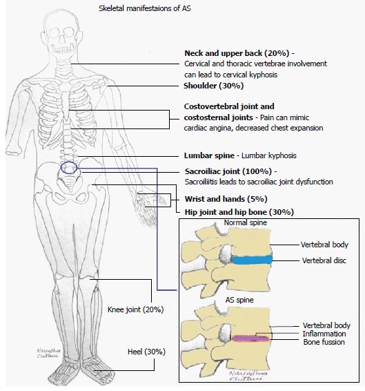
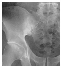
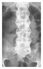
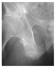
Ankylosing spondylitis (AS) is a chronic inflammatory disease that affects 1% of the general population. As one of the most severe types of spondyloarthropathy, AS affects the spinal vertebrae and sacroiliac joints, causing debilitating pain and loss of mobility. The goal of this review is to provide an overview of AS, from the pathophysiological changes that occur as the disease progresses, to genetic factors that are involved with its onset. Considering the high prevalence in the population, and the debilitating life changes that occur as a result of the disease, a strong emphasis is placed on the diagnostic imaging methods that are used to detect this condition, as well as several treatment methods that could improve the health of individuals diagnosed with AS.

Keywords: Ankylosing spondylitis; Computed tomography; Diagnosis; Magnetic resonance imaging; Treatment; Ultrasound.


Figures

Figure 1
Figure 1
Schematic of skeletal manifestations of ankylosing spondylitis. AS: Ankylosing spondylitis.
Figure 2
Figure 2
Schematic of extra-skeletal manifestations of ankylosing spondylitis. AS: Ankylosing spondylitis.
Figure 3
Figure 3
Anteroposterior pelvic X-ray shows pseudodilatation of the sacroiliac joint due to subchondral erosion and subchondral bone sclerosis.
Figure 4
Figure 4
Anteroposterior vertebral X-ray shows bilateral thickening of the syndesmophytes resulting in the appearance of the “bamboo sign” in radiographs.
Figure 5
Figure 5
Anteroposterior pelvic X-ray shows ill-defined erosions with sclerosis at the side of ligaments, and tendonitis is seen as enthesopathy at certain sites around the pelvis, at the ischial tuberosity.
Figure 6
Figure 6
Computed tomography of sacroiliac joint shows bilateral subchondral sclerosis and dentate joint contour due to bone erosion.
Figure 7
Figure 7
Color Doppler sonogram of sacroiliac joint in a patient with active sacroiliitis reveals vascularization within the posterior portion of the right sacroiliac joint.
Figure 8
Figure 8
Color and spectral waveform Doppler ultrasonogram of the ischial tuberosity in a patient with severe enthesopathy with a mean resistive index of 0.74 before treatment.
Figure 9
Figure 9
Ultrasonogram shows increased intima-media thickness of both near (0.82 mm) and far wall (0.77 mm) of common carotid artery in patients with ankylosing spondylitis.
Figure 10
Figure 10
Oblique coronal magnetic resonance imaging (short tau inversion recovery sequence) of a patient with active ankylosing spondylitis revealed bilateral high signal intensity in both the sacral and iliac components of the sacroiliac joint.

References

    1. Hukins DWL, Meakin JR. Relationship between structure and mechanical function of the tissues of the intervertebral joint. American Zoology. 2000;40:42–52.
    1. Vleeming A, Schuenke MD, Masi AT, Carreiro JE, Danneels L, Willard FH. The sacroiliac joint: an overview of its anatomy, function and potential clinical implications. J Anat. 2012;221:537–567. - PMC - PubMed
    1. Moore KL, Dalley AF, Agur AMR. Clinically Oriented Anatomy, 7th ed. PA: Lippincott Williams & Wilkins; 2014.
    1. Forst SL, Wheeler MT, Fortin JD, Vilensky JA. The sacroiliac joint: anatomy, physiology and clinical significance. Pain Physician. 2006;9:61–67. - PubMed
    1. Benjamin M, Ralphs JR. Entheses--the bony attachments of tendons and ligaments. Ital J Anat Embryol. 2001;106:151–157. - PubMed