. 2015:2015:865821.
doi: 10.1155/2015/865821. Epub 2015 Sep 7.

Didelphys Uterus: A Case Report and Review of the Literature

Affiliations

Didelphys Uterus: A Case Report and Review of the Literature

Shadi Rezai et al. Case Rep Obstet Gynecol. 2015.

Abstract

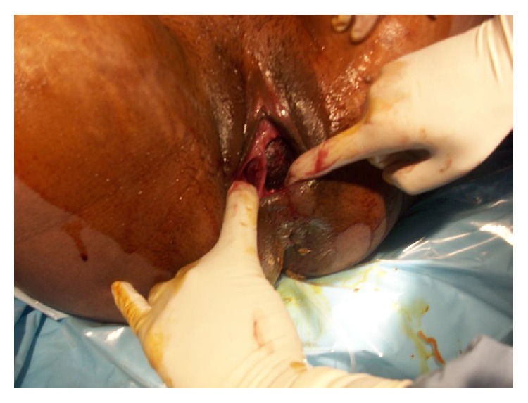
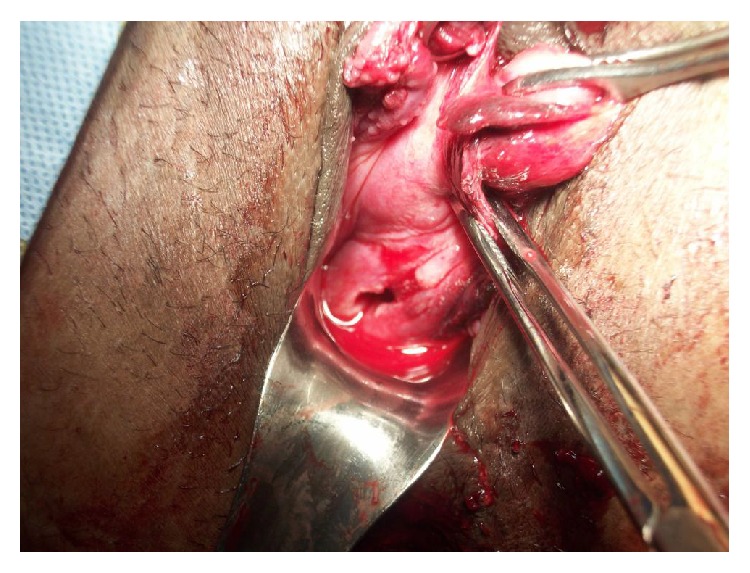
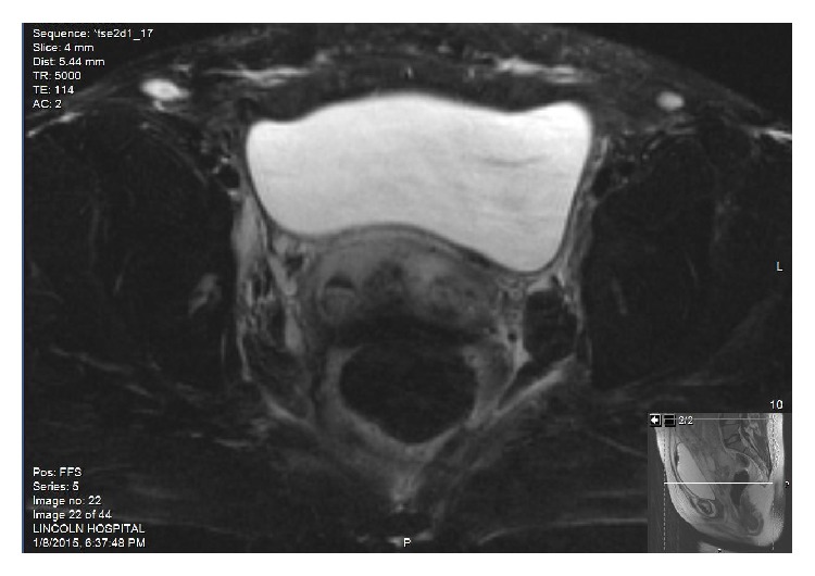
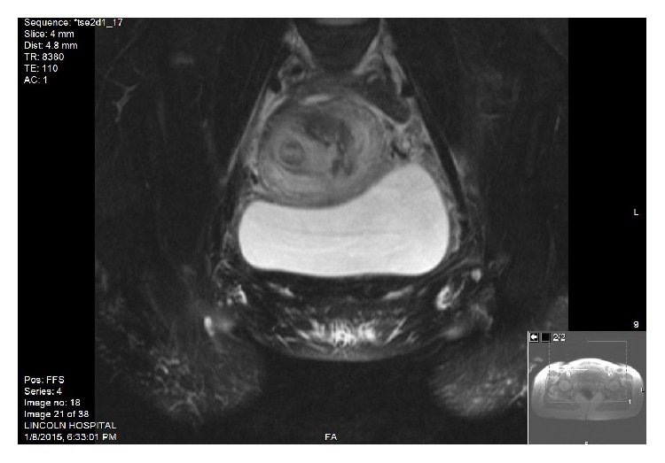
Background. Mullerian duct anomalies (MDAs) are congenital defects of the female genital system that arise from abnormal embryological development of the Mullerian ducts. A didelphys uterus, also known as a "double uterus," is one of the least common amongst MDAs. This report discusses a case of didelphys uterus that successfully conceived, carried her pregnancy to term, and delivered vaginally without any significant complications. Case. Patient is a 29-year-old G2P0010 from Bangladesh, initially came a year prior in her first pregnancy, with spontaneous abortion (SAB). Pelvic Sonogram at that time showed a diagnosis of bicornuate versus didelphys uterus. There were no renal anomalies on subsequent abdominal CT scan. Patient presented with the second pregnancy and had uncomplicated prenatal care and did not have signs of preterm labor; fetus showed appropriate growth and the pregnancy was carried in the left uterus. Patient presented at 38 4/7 wks with Premature Rupture of Membrane and underwent induction of labor with Cytotec. Antibiotics were started for chorioamnionitis. Patient had a vaginal delivery with left mediolateral episiotomy and complete tear of vaginal septum. Third stage of labor was complicated with retained placenta, which was removed manually in the operating room with total EBL of 600 cc.


Figures

Figure 1
Figure 1
Noncommunicable vaginal septum
Figure 2
Figure 2
More tear of vaginal septum as patient continues to push and fetal head descends down.
Figure 3
Figure 3
Patient was taken to operating room for removal of retained placenta; 2 separated cervices at 2 o'clock (postpartum cervix) and 7 o'clock (nonpregnant cervix) as well as completely torn vaginal septum (at 11 o'clock) are shown. Note that vaginal septum was completely destructed as fetal head delivered.
Figure 4
Figure 4
MRI of abdomen and pelvis with contrast: Series # 5, T2 axial FS: one cervix on the right and one cervix on the left, 2 separate cervices.
Figure 5
Figure 5
MRI of abdomen and pelvis with contrast: Series # 4 coronal FS (Fast): right uterus and left bulky postpartum uterus.

References

    1. Heinonen P. K. Uterus didelphys: a report of 26 cases. European Journal of Obstetrics & Gynecology and Reproductive Biology. 1984;17(5):345–350. - PubMed
    1. Heinonen P. K. Clinical implications of the didelphic uterus: long-term follow-up of 49 cases. European Journal of Obstetrics & Gynecology and Reproductive Biology. 2000;91(2):183–190. doi: 10.1016/S0301-2115(99)00259-6. - DOI - PubMed
    1. Raga F., Bauset C., Remohi J., Bonilla-Musoles F., Simón C., Pellicer A. Reproductive impact of congenital Müllerian anomalies. Human Reproduction. 1997;12(10):2277–2281. doi: 10.1093/humrep/12.10.2277. - DOI - PubMed
    1. Grimbizis G. F., Camus M., Tarlatzis B. C., Bontis J. N., Devroey P. Clinical implications of uterine malformations and hysteroscopic treatment results. Human Reproduction Update. 2001;7(2):161–174. doi: 10.1093/humupd/7.2.161. - DOI - PubMed
    1. Acién P. Reproductive performance of women with uterine malformations. Human Reproduction. 1993;8(1):122–126. - PubMed

LinkOut - more resources