

doi: 10.1155/2015/927286.
Epub 2015 Sep 7.
Anastomosing Haemangioma of the Kidney Involving a Segmental Branch of the Renal Vein
Affiliations
- PMID: 26435872
- PMCID: PMC4576015
- DOI: 10.1155/2015/927286
Item in Clipboard
Anastomosing Haemangioma of the Kidney Involving a Segmental Branch of the Renal Vein
Case Rep Surg.
2015.
Abstract
Anastomosing variant of capillary haemangioma is a rare and recently described vascular tumour with a proclivity for the genitourinary tract. Here we present the case of a 64-year-old man with incidental finding of 3.4 cm renal mass on CT who had laparoscopic nephrectomy with a good postoperative recovery. Histopathological diagnosis of anastomosing haemangioma of the kidney was made and the patient was followed up for 10 months without evidence of tumour recurrence.
Figures
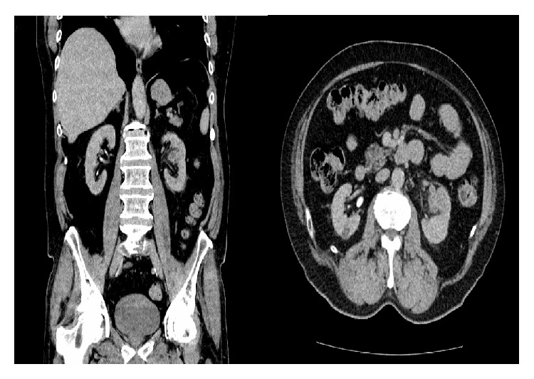
CT imaging of the mid pole solid mass of the left kidney.
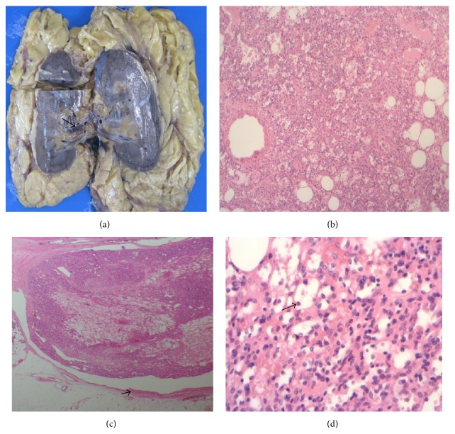
(a) Macroscopic image of the tumour in the renal hilum with involvement of the branch of a segmental renal vein (arrowed). (b) 40x H&E showing the tumour with sieve-like pattern. (c) 20x H&E demonstrating the involvement of the tributary of a segmental renal vein (vessel wall arrowed). (d) 200x H&E showing round to oval vasoformative endothelial cells with hobnail appearance (arrowed).
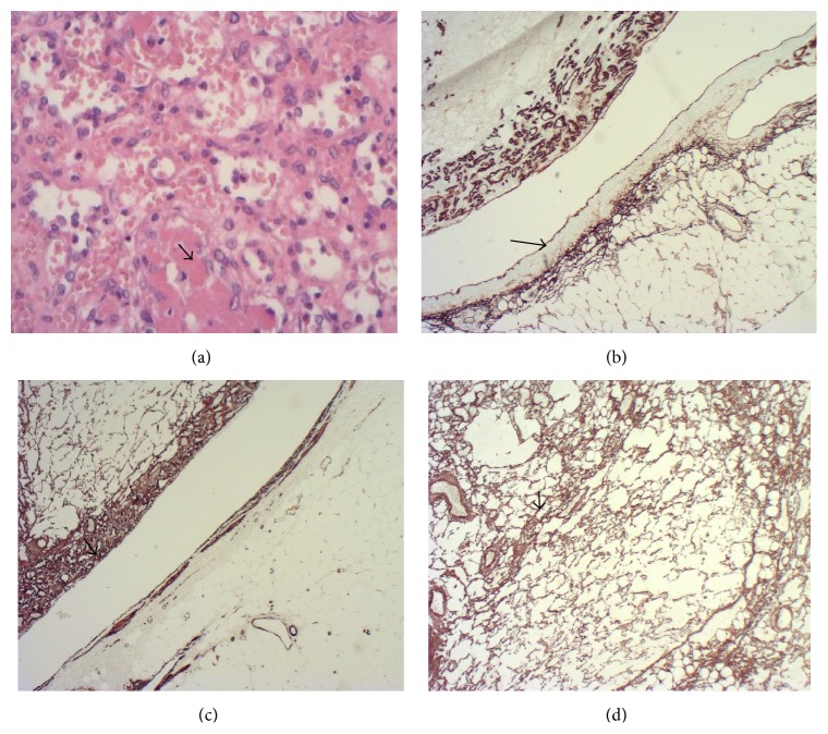
(a) 200x H&E showing fibrin thrombi within some vascular spaces. (b) showing CD34 immunohistochemistry staining the endothelium in the tumour as well as the segmental renal vein branch (arrowed) (×20). (c) demonstrating smooth muscle actin immunohistochemistry staining the tumour pericytes (arrowed) and vessel wall (×20). (d) showing CD34 immunohistochemistry staining supporting pericytes (×40).
References
-
- Kryvenko O. N., Gupta N. S., Meier F. A., Lee M. W., Epstein J. I. Anastomosing hemangioma of the genitourinary system: eight cases in the kidney and ovary with immunohistochemical and ultrastructural analysis. American Journal of Clinical Pathology. 2011;136(3):450–457. doi: 10.1309/ajcpjpw34qcqytmt. - DOI - PubMed
LinkOut - more resources
Full Text Sources
Other Literature Sources
