

Analyzing bone remodeling patterns after total hip arthroplasty using quantitative computed tomography and patient-specific 3D computational models
- PMID: 26435921
- PMCID: PMC4559985
- DOI: 10.3978/j.issn.2223-4292.2015.08.03
Analyzing bone remodeling patterns after total hip arthroplasty using quantitative computed tomography and patient-specific 3D computational models
Abstract
Background: Computational models in the form of finite element analysis technique that incorporates bone remodeling theories along with DEXA scans has been extensively used in predicting bone remodeling patterns around the implant. However, majority of such studies used generic models. Therefore, the aim of this study is to develop patient-specific finite element models of total hip replacement patients using their quantitative computed tomography (QCT) scans and accurately analyse bone remodelling patterns after total hip arthroplasty (THA).
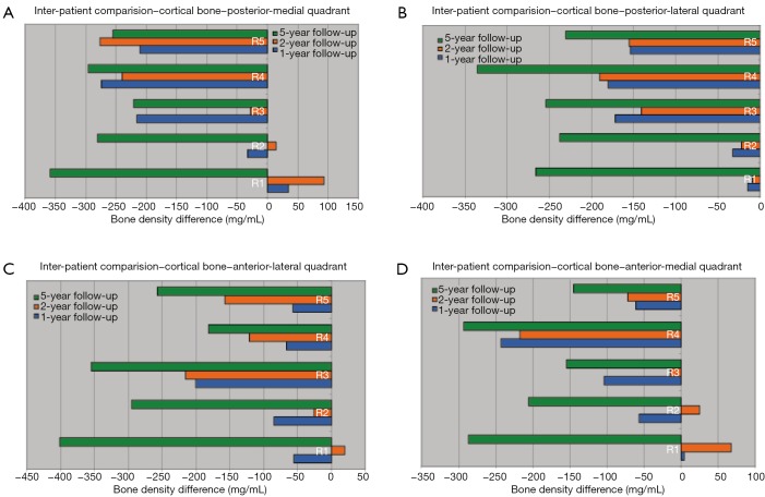
Methods: Patient-specific finite element models have been generated using the patients' QCT scans from a previous clinical follow-up study. The femur was divided into five regions in proximal-distal direction and then further divided into four quadrants for detailed analysis of bone remodeling patterns. Two types of analysis were performed-inter-patient and intra patient to compare them and then the resulting bone remodeling patterns were quantitatively analyzed.
Results: Our results show that cortical bone density decrease is higher in diaphyseal region over time and the cancellous bone density decreases significantly in metaphyseal region over time. In metaphyseal region, posterior-medial (P-M) quadrant showed high bone loss while diaphyseal regions show high bone loss in anterior-lateral (A-L) quadrant.
Conclusions: Our study demonstrated that combining QCT with 3D patient-specific models has the ability of monitoring bone density change patterns after THA in much finer details. Future studies include using these findings for the development of a bone remodelling algorithm capable of predicting surgical outcomes for THA patients.
Keywords: Quantitative computed tomography (QCT); bone remodeling; finite element analysis; total hip arthroplasty (THA).
Conflict of interest statement
Figures


Similar articles
-
Quantitative CT with finite element analysis: towards a predictive tool for bone remodelling around an uncemented tapered stem.Int Orthop. 2012 Jul;36(7):1363-9. doi: 10.1007/s00264-012-1513-x. Epub 2012 Apr 12. Int Orthop. 2012. PMID: 22527334 Free PMC article.
-
Finite element model of a novel short stemmed total hip arthroplasty implant developed from cross sectional CT scans.Technol Health Care. 2013;21(5):493-500. Technol Health Care. 2013. PMID: 24252858
-
Assessment of total hip arthroplasty by means of computed tomography 3D models and fracture risk evaluation.Artif Organs. 2013 Jun;37(6):567-73. doi: 10.1111/aor.12033. Epub 2013 Apr 2. Artif Organs. 2013. PMID: 23550540
-
Quantitative computer-assisted osteodensitometry in total hip arthroplasty.Int Orthop. 2007 Aug;31(4):431-8. doi: 10.1007/s00264-006-0257-x. Epub 2006 Oct 17. Int Orthop. 2007. PMID: 17043862 Free PMC article. Review.
-
Computer-assisted osteodensitometry following total hip arthroplasty.Expert Rev Med Devices. 2006 Nov;3(6):763-8. doi: 10.1586/17434440.3.6.763. Expert Rev Med Devices. 2006. PMID: 17280541 Review.
Cited by
-
Evaluation of peri-prosthetic radiolucent lines surrounding the cementless femoral stem using digital tomosynthesis with metal artifact reduction: a cadaveric study in comparison with radiography and computed tomography.Quant Imaging Med Surg. 2020 Sep;10(9):1786-1800. doi: 10.21037/qims-19-1018. Quant Imaging Med Surg. 2020. PMID: 32879857 Free PMC article.
-
High periprosthetic bone mineral density measured in immediate postoperative period may not guarantee less periprosthetic bone loss in the proximal femur after cementless total hip arthroplasty - A retrospective study.Arthroplasty. 2020 Jan 23;2(1):2. doi: 10.1186/s42836-020-0023-3. Arthroplasty. 2020. PMID: 35236466 Free PMC article.
-
Stress-Strain State Investigation and Ultimate Load on Femoral Implants Based on S-Type Ti6Al4V Titanium Alloy.J Funct Biomater. 2025 May 19;16(5):187. doi: 10.3390/jfb16050187. J Funct Biomater. 2025. PMID: 40422851 Free PMC article.
-
Changes in Periprosthetic Bone Mineral Density Following Arthroplasty: An In-Depth Review and Current Perspectives.Curr Osteoporos Rep. 2025 Jun 27;23(1):30. doi: 10.1007/s11914-025-00921-6. Curr Osteoporos Rep. 2025. PMID: 40576727 Review.
-
Quantitative Computed Tomography (QCT) derived Bone Mineral Density (BMD) in finite element studies: a review of the literature.J Exp Orthop. 2016 Dec;3(1):36. doi: 10.1186/s40634-016-0072-2. Epub 2016 Dec 9. J Exp Orthop. 2016. PMID: 27943224 Free PMC article. Review.
References
-
- Bobyn JD, Engh CA, Glassman AH. Histologic analysis of a retrieved microporous-coated femoral prosthesis. A seven-year case report. Clin Orthop Relat Res 1987;(224):303-10. - PubMed
-
- Engh CA, Bobyn JD, Glassman AH. Porous-coated hip replacement. The factors governing bone ingrowth, stress shielding, and clinical results. J Bone Joint Surg Br 1987;69:45-55. - PubMed
-
- Rüegsegger P, Koller B, Müller R. A microtomographic system for the nondestructive evaluation of bone architecture. Calcif Tissue Int 1996;58:24-9. - PubMed
-
- Cowin SC. Bone stress adaptation models. J Biomech Eng 1993;115:528-33. - PubMed
-
- Huiskes R, van Rietbergen B. Preclinical testing of total hip stems. The effects of coating placement. Clin Orthop Relat Res 1995;(319):64-76. - PubMed
LinkOut - more resources
Full Text Sources