Review
. 2015 Aug;5(4):592-602.
doi: 10.3978/j.issn.2223-4292.2015.08.01.

Identifying osteoporotic vertebral fracture

Affiliations
Review

Identifying osteoporotic vertebral fracture

James F Griffith. Quant Imaging Med Surg. 2015 Aug.

Abstract

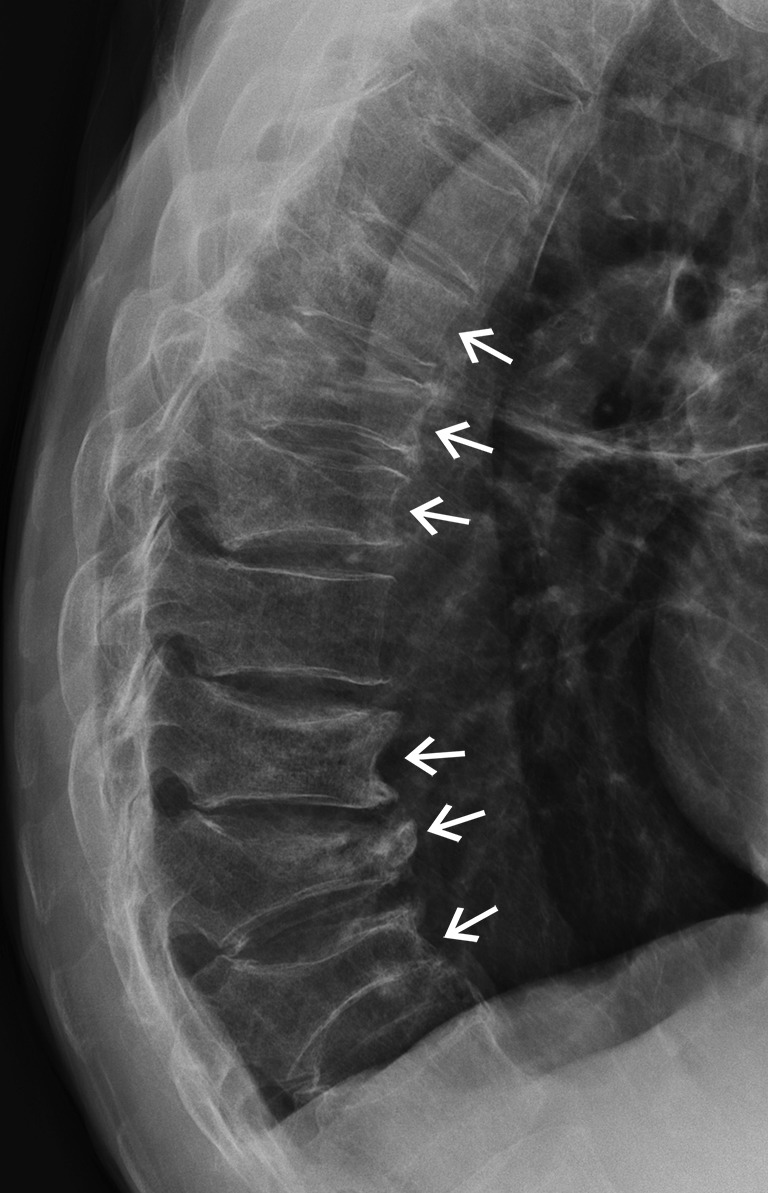
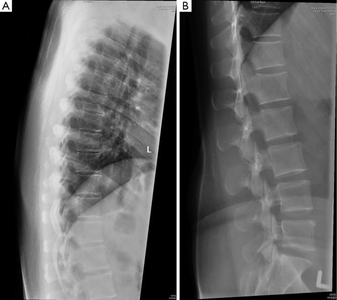
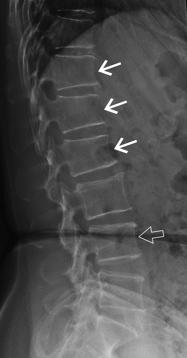
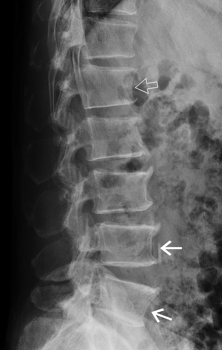
Osteoporosis per se is not a harmful disease. It is the sequela of osteoporosis and most particularly the occurrence of osteoporotic fracture that makes osteoporosis a serious medical condition. All of the preventative measures, investigations, treatment and research into osteoporosis have one primary goal and that is to prevent the occurrence of osteoporotic fracture. Vertebral fracture is by far and away the most prevalent osteoporotic fracture. The significance and diagnosis of vertebral fracture are discussed in this article.

Keywords: Vertebral fracture; computed tomography (CT); magnetic resonance imaging (MRI); metastases; osteoporosis; radiography.


Conflict of interest statement

Conflicts of Interest: The author has no conflicts of interest to declare.

Figures

Figure 1
Figure 1
Lateral radiograph showing vertebral fracture cascade with fractures of many contiguous or near contiguous vertebrae.
Figure 2
Figure 2
Lateral radiographs showing (A) normal thoracic and (B) normal lumbar vertebrae.
Figure 3
Figure 3
Difficult call. Distinguishing between physiological wedging of the T12, L1 and L2 vertebral bodies (arrows) and a mild fracture is difficult in this case. Anterior vertebral body height is not quite reduced to >20% of what you would normally expect it to be (given expected physiological wedging at this level). There is also short vertebral height of the L4 vertebral body (open arrow) in that the height is not reduced to more than 20% of what you would normally expect it to be at this level.
Figure 4
Figure 4
There is short vertebral height of all the lumbar vertebrae particularly L4 and L5 (arrows). There is also physiological wedging of L1 (open arrow).
Figure 5
Figure 5
Schematic diagram showing endplate impressions caused by (A) Scheuermann’s disease; (B) Schmorl’s node and (C) Cupid’s bow deformity.
Figure 6
Figure 6
There is a severe osteoporotic fracture of the T12 vertebral body (open arrow) with a mild osteoporotic fracture of the L1 vertebral body (arrow). There is a lumbar scoliosis with obliquity of the lower three lumbar vertebrae (*).
Figure 7
Figure 7
Schematic diagram of Genant semi-quantitative analysis of vertebral fracture severity.
Figure 8
Figure 8
Lateral thoracic spine radiograph. There is a severe fracture of the T11 vertebral body (arrow) with a moderate fracture of the T4 vertebral body (thick arrow).
Figure 9
Figure 9
Schematic diagram showing examples of reference point placement for quantitative morphometry.
Figure 10
Figure 10
Vertebral fracture assessment by DXA showing mild fracture of L1 vertebral body. DXA, dual energy X-ray absorptiometry.
Figure 11
Figure 11
T2-weighted sagittal MRI showing typical osteoporotic-type fracture of T12 vertebral body with preservation of some marrow fat and fluid filled cavity within vertebral body (closed arrow). There is a chronic fracture of the L3 vertebral body with fat filling the marrow cavity (open arrow). MRI, magnetic resonance imaging.

Similar articles

Cited by

References

    1. Grigoryan M, Guermazi A, Roemer FW, Delmas PD, Genant HK. Recognizing and reporting osteoporotic vertebral fractures. Eur Spine J 2003;12:S104-12. - PMC - PubMed
    1. Cooper C, Atkinson EJ, O'Fallon WM, Melton LJ, 3rd. Incidence of clinically diagnosed vertebral fractures: a population-based study in Rochester, Minnesota, 1985-1989. J Bone Miner Res 1992;7:221-7. - PubMed
    1. Lips P, van Schoor NM. Quality of life in patients with osteoporosis. Osteoporos Int 2005;16:447-55. - PubMed
    1. Kado DM, Browner WS, Palermo L, Nevitt MC, Genant HK, Cummings SR. Vertebral fractures and mortality in older women: a prospective study. Study of Osteoporotic Fractures Research Group. Arch Intern Med 1999;159:1215-20. - PubMed
    1. Siris ES, Miller PD, Barrett-Connor E, Faulkner KG, Wehren LE, Abbott TA, Berger ML, Santora AC, Sherwood LM. Identification and fracture outcomes of undiagnosed low bone mineral density in postmenopausal women: results from the National Osteoporosis Risk Assessment. JAMA 2001;286:2815-22. - PubMed