. 2015 Jun;26(1-2):70-6.
doi: 10.1007/s13337-015-0253-0. Epub 2015 May 23.

Characterization of cytopathogenicity of classical swine fever virus isolate induced by Newcastle disease virus

Affiliations

Characterization of cytopathogenicity of classical swine fever virus isolate induced by Newcastle disease virus

S D Raut et al. Virusdisease. 2015 Jun.

Abstract

Classical swine fever virus (CSFV), the causative agent of classical swine fever, belongs to the family Flaviviridae and genus Pestivirus. Some pestiviruses exhibit cytopathic effect in cell culture but exact phenomenon is unknown. Over expression of NS2-3 gene, presence of defective interfering particle and exaltation of Newcastle disease virus (END) phenomenon could be the reasons of cytopathogenicity. In the present study, a CSFV isolate exhibiting cytopathic effect (CPE) in Madin-Darby Canine Kidney (MDCK) cell line was characterized. To characterize cytopathogenicity of such isolate, END test was carried out. Interference of Newcastle disease virus (NDV) in MDCK adapted CSFV was confirmed by RT-PCR and virus neutralization test. Absence of CPE and NDV specific nucleic acid after neutralization confirmed the induction of CPE by NDV. Further, identity of the CSFV isolate in MDCK cell line by immunoperoxidase test, immunoblotting and RT-PCR post NDV neutralization established the virus replication without CPE (non-cytopathic isolate). Findings suggest that, there could be a chance of mixed infection of both CSFV and NDV in the piglet from which the sample was collected for virus isolation.

Keywords: CSF; Cytopathogenicity; END phenomenon; MDCK; VNT; Virus.


Figures
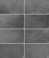
Fig. 1
Fig. 1
ah CPE in Madin-Darby Canine Kidney (MDCK) cell-line infected with cytopathic CSFV (P25): a, c, e and g: Healthy cells (1st to 4th dpi); b look alike healthy cell (1st dpi); d rounding (2nd dpi); f clumping (3rd dpi) and h detachment (4th dpi) of cells
Fig. 2
Fig. 2
a, b RT-PCR targeting 5′UTR and E2 genes of representative passages (P25, 30 and 35) of CSFV isolate adapted to MDCK cells: 5′UTR (a) and E2 (b) based amplification of representative passages of CSFV isolate; LVV Lapinised vaccine virus (positive control), M 100 bp DNA ladder plus Marker (MBI, Fermentas), NTC Non template control
Fig. 3
Fig. 3
Presence of NDV (356 bp) in CSFV isolate (P 25–35): M 100 bp ladder, L1 P-25, L2 P-30, L3 P-35, L4 Positive control, NTC Non-template control
Fig. 4
Fig. 4
a, b Post neutralization RT-PCR of NDV (a) and CSFV (b). NDV couldn’t be detected up to 1:16 dilution, whereas CSFV gene amplified in the same dilution. 1:8 to 1:64: virus-serum dilutions used for neutralization, M 100 bp DNA ladder, VC Virus control, +VeC positive control, NTC non-template control
Fig. 5
Fig. 5
Post-neutralization Western blot analysis of CSFV immunogenic protein (E2) using MAb.: M Prestained protein marker, Lane 1 CSFV infected MDCK cell supernatant, Lane 2 mock infected MDCK cell supernatant, Lane 3 mock infected MDCK cell-lysate, Lane 4 CSFV infected MDCK cell-lysate
Fig. 6
Fig. 6
Post-neutralization IPT staining of MDCK cells infected with CSFV: IPT showing dark brawn colour in infected and no stain in mock infected cells by MAb (ai, ii) directed against CSFV E2 (1:40) and CSFV antiserum (1:20) (bi, ii), antimouse conjugate (1:200), 3-Amino-9-Ethyl Carbazole (AEC) as chromogen and 3 % H2O2 as substrate

Similar articles

References

    1. Aoki H, Ishikawa K, Sakoda Y, Sekiguchi H, Kodama M, Suzuki S, Fukusho A. Characterization of classical swine fever virus associated with defective interfering particles containing a cytopathogenic subgenomic RNA isolated from wild boar. J Vet Med Sci. 2001;63:751–758. doi: 10.1292/jvms.63.751. - DOI - PubMed
    1. Aoki H, Ishikawa K, Sekiguchi H, Suzuki S, Fukusho A. Pathogenicity and kinetics of virus propagation in swine infected with the cytopathogenic classical swine fever virus containing defective interfering particles. Arch Virol. 2003;148:297–310. doi: 10.1007/s00705-002-0907-2. - DOI - PubMed
    1. Cheng LF, Ke MF, Zheng T, Huang Y, Li WY, Shi SH. Isolation and identification of Newcastle disease virus from swine. Chin J Vet Sci Tech. 2004–12. http://en.cnki.com.cn/Article_en/CJFDTOTAL-ZGSY200412020.htm.
    1. Chomczynski P, Sacchi N. Single-step method of RNA isolation by guanidinium thiocyanate-phenol chloroform extraction. Anal Biochem. 1987;162:156–159. doi: 10.1016/0003-2697(87)90021-2. - DOI - PubMed
    1. Devereux J, Haeberli P, Smithies O. A comprehensive set of sequence analysis programsfor the VAX. Nucleic Acids Res. 1984;12:387–395. doi: 10.1093/nar/12.1Part1.387. - DOI - PMC - PubMed

LinkOut - more resources