. 2015 Nov;17(11):1412-21.
doi: 10.1038/ncb3250. Epub 2015 Oct 5.

Integrin endosomal signalling suppresses anoikis

Affiliations

Integrin endosomal signalling suppresses anoikis

Jonna Alanko et al. Nat Cell Biol. 2015 Nov.

Abstract

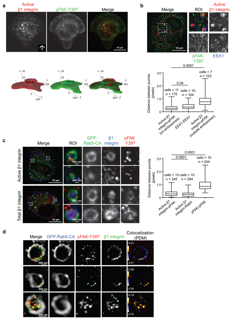
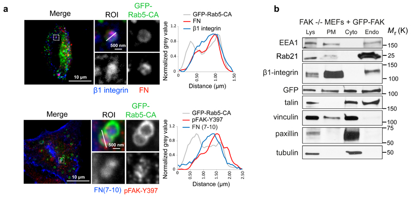
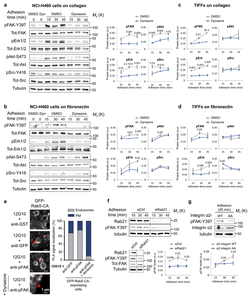
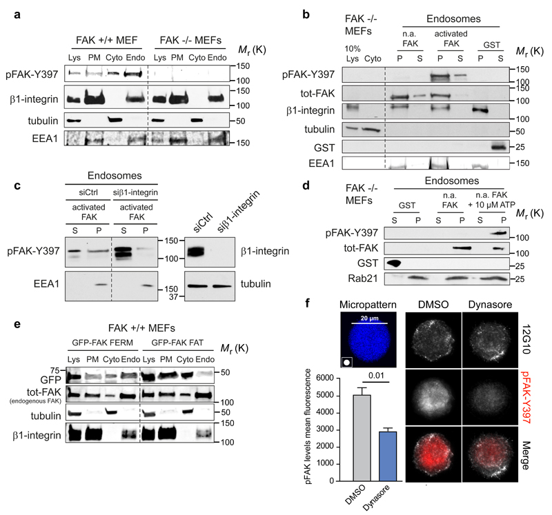
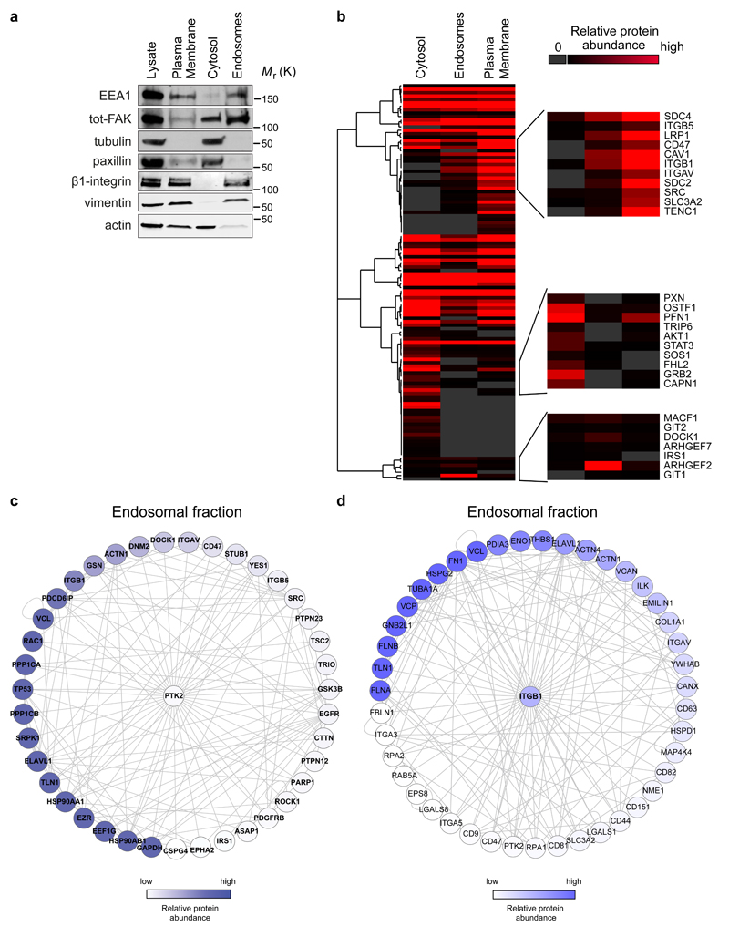
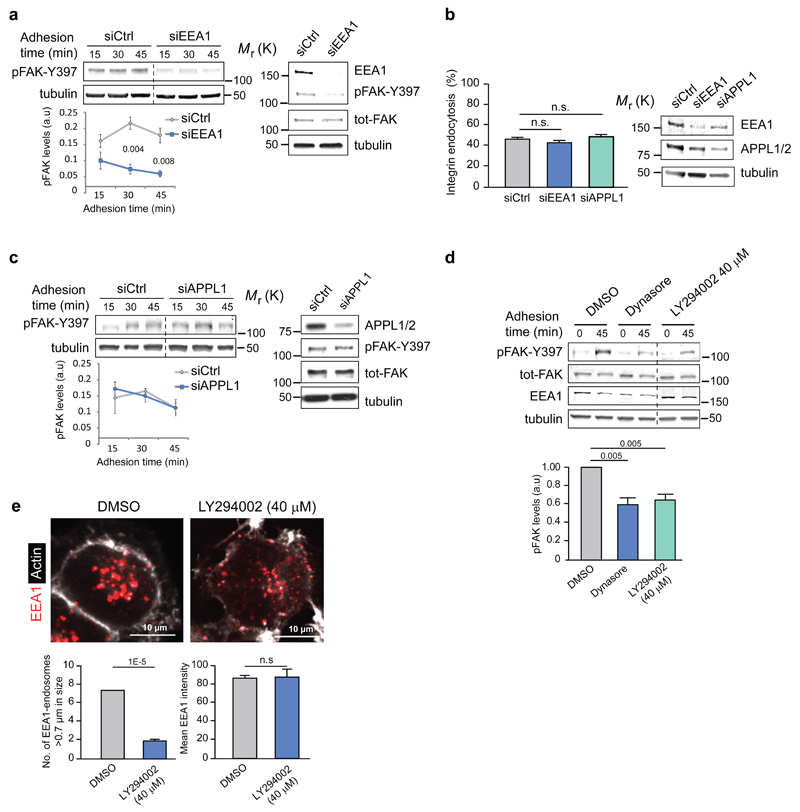
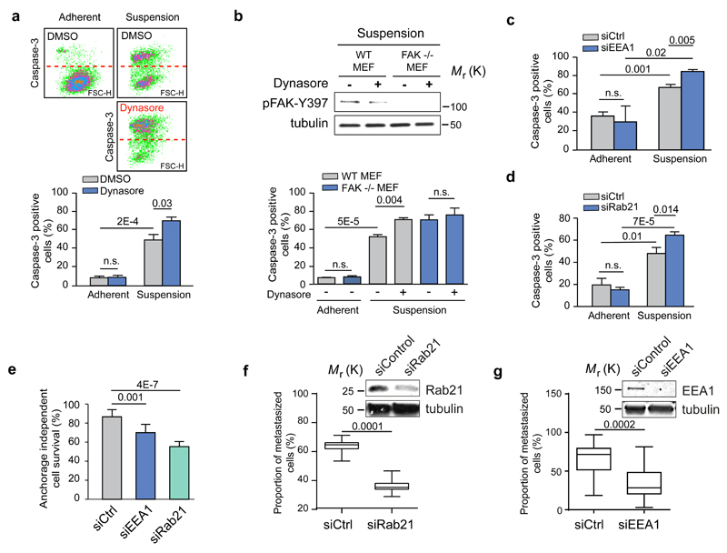
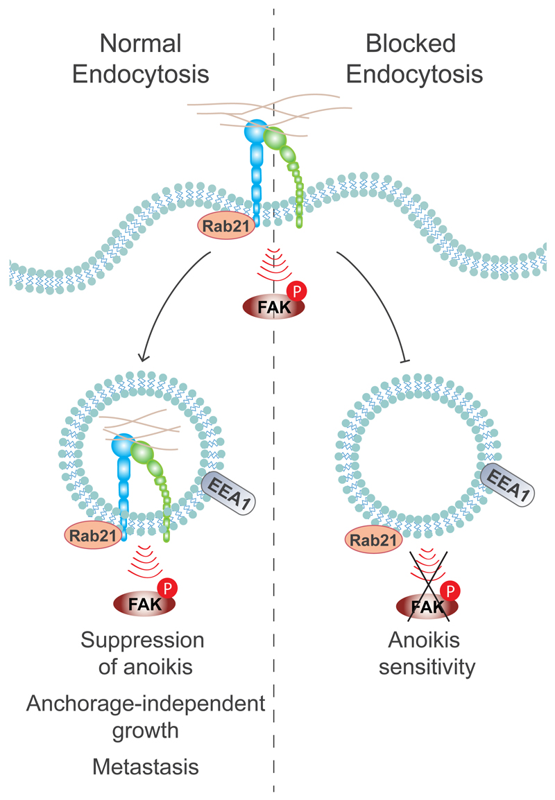
Integrin-containing focal adhesions transmit extracellular signals across the plasma membrane to modulate cell adhesion, signalling and survival. Although integrins are known to undergo continuous endo/exocytic traffic, the potential impact of endocytic traffic on integrin-induced signals is unknown. Here, we demonstrate that integrin signalling is not restricted to cell-ECM adhesions and identify an endosomal signalling platform that supports integrin signalling away from the plasma membrane. We show that active focal adhesion kinase (FAK), an established marker of integrin-ECM downstream signalling, localizes with active integrins on endosomes. Integrin endocytosis positively regulates adhesion-induced FAK activation, which is early endosome antigen-1 and small GTPase Rab21 dependent. FAK binds directly to purified endosomes and becomes activated on them, suggesting a role for endocytosis in enhancing distinct integrin downstream signalling events. Finally, endosomal integrin signalling contributes to cancer-related processes such as anoikis resistance, anchorage independence and metastasis.


Conflict of interest statement

Authors declare no conflict of interests.

Figures

Figure 1
Figure 1. pFAK-Y397 localizes to endosomes together with β1-integrin.
a, Representative images and 3D probabilistic density maps of active β1-integrin and pFAK-Y397 localisation in MDA-MB-231 cells plated on crossbow-shaped fibronectin-coated micropatterns (24 cells assessed in three independent experiments). b, Representative confocal images of endogenous active β1-integrin, pFAK and EEA1 staining in MDA-MB-231 cells and box plot of the distance between adjacent puncta of active β1-integrin and pFAK in or outside the EEA1-positive endosomes (in pixels) (box plots show the 25th–75th percentiles delineated by the upper and lower limits of the box; the median is shown by the horizontal line inside the box. Whiskers indicate maxima and minima). n= the number of active β1-integrin-pFAK and EEA1-EEA1 doublets (indicated in the figure) analysed from multiple cells (numbers indicated in the figure) from three independent experiments are indicated. c, Representative confocal images of pFAK and total or active β1-integrin staining in GFP-Rab5-CA-expressing TIFFs adhering to fibronectin (45 min). Box plot of the distance between adjacent puncta of active β1-integrin and pFAK or Rab5 in GFP-Rab5-positive endosomes and between pFAK and pFAK outside the endosomes (in pixels) (box plots show the 25th–75th percentiles delineated by the upper and lower limits of the box; the median is shown by the horizontal line inside the box. Whiskers indicate maxima and minima). Numbers of cells and of active β1-integrin-pFAK, active β1-integrin-Rab5 and pFAK-pFAK doublets (n) analysed from three independent experiments are indicated. d, Super-resolution STED images of active β1-integrin and pFAK-Y397 on single GFP-Rab5-CA endosomes in TIFFs adhering to fibronectin (45 min). Images displaying PDM (product of the differences from the mean) values were generated to visualize the colocalization between β1-integrin and pFAK-Y397. ROI: region of interest. Mann-Whitney test P values are provided.
Figure 2
Figure 2. pFAK-Y397 localizes to endosomes together with fibronectin.
a, Active β1 integrin, pFAK and fibronectin localisation in GFP-Rab5-CA-expressing TIFFs plated on fibronectin (top panel) or incubated with 647-labelled fibronectin fragment (FN(7-10)) for 45 min (bottom panel). Fluorescence intensity profiles (normalized grey value) across representative endosomes (solid lines) are illustrated. b, Subcellular fractionation of FAK -/- MEFs re-expressing GFP-FAK and representative immunoblots from more than three independent experiments. Lys: cell lysate; PM: plasma membrane; Cyto: cytosol; Endo: Endosomal fraction; ROI: region of interest. Uncropped images of blots are shown in supplementary figure 9.
Figure 3
Figure 3. Inhibition of integrin endocytosis attenuates integrin signalling.
a-d, Analysis of kinase activity in NCI-H460 cells (a, b) and TIFFs (c, d) ± dynasore plated on collagen (a, c) or fibronectin (b, d) (mean ± SEM, a, n=5 independent experiments; b-d, n=3 independent experiments). e, In-situ PLA signal (red dots) representing colocalisation between 12G10 (active integrin antibody) and the indicated antibodies in GFP-Rab5-CA-expressing cells (10 cells assessed from two independent experiments). f, Representative immunoblot and quantification of pFAK protein levels in Rab21- or control-silenced NCI-H460 cells plated on collagen for the indicated times (mean ± SEM, n=3 independent experiments). g, Representative immunoblot and quantification of pFAK in CHO cells expressing α2-integrin wild-type (WT) or Rab21-binding deficient integrin α2-AA-mutant and plated on collagen for 45 min (mean ± SEM, n=3 independent experiments). a.u: arbitrary units. Student's two-tailed unpaired t-test P values are provided and statistics source data can be found in Supplementary Table 2. Uncropped images of blots are shown in supplementary figure 9.
Figure 4
Figure 4. FAK recruitment and activation on integrin-positive endosomes
a, Representative immunoblot of subcellular fractionation of FAK+/+ and FAK-/- MEFs (five independent experiments). b, Representative immunoblot of recombinant FAK (phosphorylated/activated or non-activated: n.a) recruitment to insoluble endosomal pellet (P) and soluble supernatant (S) fractions isolated from FAK -/- MEFs (five independent experiments). c, Representative immunoblot analysing the recruitment of recombinant FAK to purified endosomes derived from either control- or β1-integrin-silenced FAK -/- MEFs (five independent experiments). d, Representative immunoblot analysing the activation of recombinant FAK in purified endosome fractions derived from FAK -/- MEFs in the presence or absence of 10 µM ATP (five independent experiments). e, Subcellular fractionation of FAK+/+ MEFs transfected with GFP-FAK FAT or GFP-FAK FERM. Shown are representative immunoblots of GFP-FAK fragments and endogenous FAK (total-FAK) from two independent experiments. f, MDA-MB-231 cells plated on 20 μm round fibronectin-coated micropatterns (45 min) ± dynasore. Representative maximum projections and quantification of pFAK (mean fluorescence ± SEM, n=3 independent experiments, 10 cells analysed / experiment). Lys: cell lysate; PM: plasma membrane; Cyto: cytosol; Endo: Endosomal fraction. Student's two-tailed unpaired t-test P =0.01. Uncropped images of blots are shown in supplementary figure 9.
Figure 5
Figure 5. Endosomal proteome.
a, Representative western blot validation of fractionated samples analysed by mass spectrometry. b-d, The threshold for protein identification was set at a minimum of 3 spectral counts with at least 2 unique peptides. Altogether, 2021, 1667 and 2006 proteins were detected in the cytosolic, endosomal and plasma membrane fractions, respectively. b, Hierarchical clustering of Geiger adhesome proteins identified in the cytoplasmic, plasma membrane and endosomal fractions. Examples of proteins detected in multiple clusters are displayed on the right hand side (two independent experiments). c-d, Known FAK (PTK2) (c) and β1-integrin (ITGB1) (d) interacting proteins identified in the mass spectrometric analysis of purified endosomal fractions in FAK +/+ MEFs. Proteins were mapped onto a human protein-protein interaction network. Each node represents a protein (labelled with gene name) and each edge represents a reported interaction between two proteins. The nodes are coloured according to protein abundance (two independent experiments). Uncropped images of blots are shown in supplementary figure 9.
Figure 6
Figure 6. Integrin signalling is EEA-1 dependent.
a, c, Representative blots and quantification of pFAK protein levels following EEA1 (a) or APPL1 (c) silencing in NCI-H460 cells plated on collagen (mean ± SEM, n=3 independent experiments). b, Integrin endocytosis in EEA1- or APPL1-silenced NCI-H460 cells plated on collagen (30 min) using active β1-integrin (9EG7) antibody (proportion of cytoplasmic/total staining, mean ± SEM, n=43 cells pooled from three independent experiments). d, Representative immunoblot and quantification of pFAK protein levels in NCI-H460 cells plated on collagen ± dynasore or PI3K- inhibitor LY294002 (mean ± SEM, n=3 independent experiments). e, Representative confocal images of EEA1 and actin staining in NCI-H460 cells ± PI3K inhibitor (LY294002 40 μM) and quantification of the number of EEA1-endosomes larger than 0.7 μm and the total mean EEA1 intensity (mean ± SEM, n = 16 cells per condition pooled from two independent experiments). Student's two-tailed unpaired t-test P values are provided and statistics source data can be found in Supplementary Table 2. Uncropped images of blots are shown in Supplementary Figure 9.
Figure 7
Figure 7. Integrin endosomal signalling and anoikis sensitivity are Rab21- and EEA1- dependent.
a-d, Quantification of caspase-3 positive (FL1) apoptotic cells in serum-starved TIFFs and representative dot blots (a), in FAK-/- or FAK+/+ WT MEFs ± dynasore (b) and following EEA1 (c) or Rab21 (d) silencing in TIFFs (mean fluorescence ± SEM, n=3 independent experiments). e, Quantification of anchorage-independent survival of MDA-MB-231 cells following EEA1 or Rab21 silencing (mean ± s.d., n=3 independent experiments). a-e, Student's two-tailed unpaired t-test P values are provided. f-g, MDA-MB-231 cells transfected with siCtrl and siRab21 (f) or siCtrl and siEEA1 (g) were fluorescently labelled with green or far-red cell trackers and coinjected 1:1 into the tail vein of mice. The proportion of extravasated cells was analysed by flow cytometry 48 hr after coinjection and is represented as a percentage of total extravasated cells in the lung (box plots show the 25th–75th percentiles delineated by the upper and lower limits of the box; the median is shown by the horizontal line inside the box. Whiskers indicate maxima and minima; siRab21: n = 15 mice from one experiment, siEEA1: n=10 & 15 mice pooled from two independent experiments). Student’s two-tailed unpaired t-test P values are provided. Uncropped images of blots are shown in supplementary figure 9. Statistics source data can be found in Supplementary Table 2.
Figure 8
Figure 8. Conclusion model: the role of adhesion-induced integrin endosomal signalling in the regulation of anoikis.
The full activation of the ECM-induced integrin pFAK-Y397 signal is dependent on Rab21-mediated integrin endocytosis to EEA1-containing early endosomes, and this is ultimately required for suppression of anoikis and anchorage-independent growth. Blocked integrin endocytosis leads to reduced FAK activation and to increased anoikis sensitivity.

Comment in

References

    1. Harburger DS, Calderwood DA. Integrin signalling at a glance. J Cell Sci. 2009;122:159–163. - PMC - PubMed
    1. Winograd-Katz SE, Fassler R, Geiger B, Legate KR. The integrin adhesome: from genes and proteins to human disease. Nat Rev Mol Cell Biol. 2014;15:273–288. - PubMed
    1. Zaidel-Bar R, Itzkovitz S, Ma'ayan A, Iyengar R, Geiger B. Functional atlas of the integrin adhesome. Nat Cell Biol. 2007;9:858–867. - PMC - PubMed
    1. De Franceschi N, Hamidi H, Alanko J, Sahgal P, Ivaska J. Integrin traffic - the update. J Cell Sci. 2015;128:839–852. - PMC - PubMed
    1. Ceresa BP, Schmid SL. Regulation of signal transduction by endocytosis. Curr Opin Cell Biol. 2000;12:204–210. - PubMed

Publication types

MeSH terms