Review
. 2015:31:83-108.
doi: 10.1146/annurev-cellbio-100814-125438. Epub 2015 Sep 30.

Mechanism and regulation of cytoplasmic dynein

Affiliations
Review

Mechanism and regulation of cytoplasmic dynein

Michael A Cianfrocco et al. Annu Rev Cell Dev Biol. 2015.

Abstract

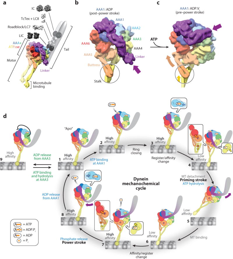
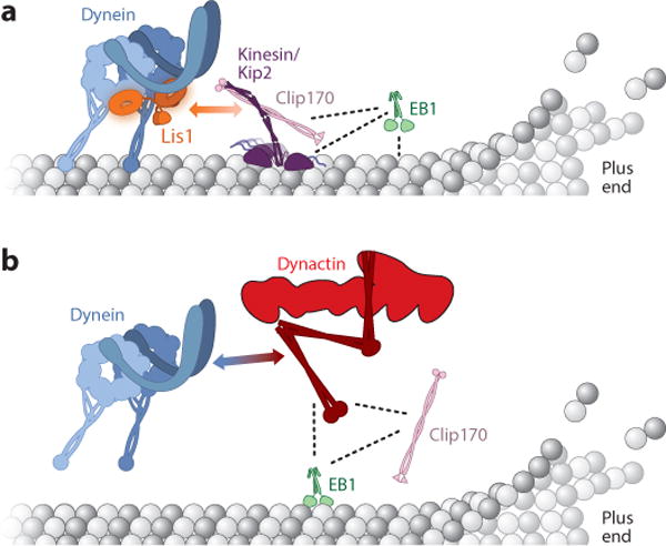
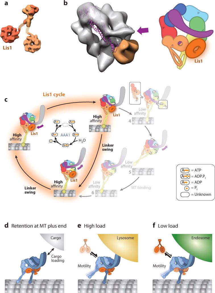
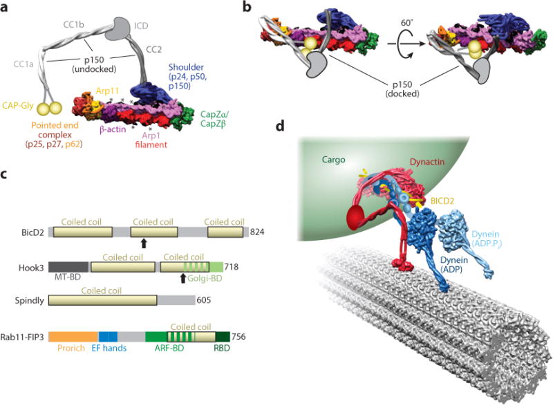
Until recently, dynein was the least understood of the cytoskeletal motors. However, a wealth of new structural, mechanistic, and cell biological data is shedding light on how this complicated minus-end-directed, microtubule-based motor works. Cytoplasmic dynein-1 performs a wide array of functions in most eukaryotes, both in interphase, in which it transports organelles, proteins, mRNAs, and viruses, and in mitosis and meiosis. Mutations in dynein or its regulators are linked to neurodevelopmental and neurodegenerative diseases. Here, we begin by providing a synthesis of recent data to describe the current model of dynein's mechanochemical cycle. Next, we discuss regulators of dynein, with particular focus on those that directly interact with the motor to modulate its recruitment to microtubules, initiate cargo transport, or activate minus-end-directed motility.

Keywords: Lis1; Nudel/NudE; cytoskeleton; dynactin; microtubule; transport.


Figures

Figure 1
Figure 1
Dynein structure and mechanochemical cycle. (a) Structure of the cytoplasmic dynein complex. The motor domain contains the AAA+ (ATPases Associated with various cellular Activities) domains, the microtubule (MT)-binding domain (MTBD), and the linker. The N-terminal tail domain binds the intermediate (IC), light intermediate (LIC), and light chains (LC). We built the structure of the motor domain by combining entries 3VKG and 4RH7 from the Protein Data Bank (PDB) and displaying the model as a density at a resolution of 10 Å. (b) Structure of the dynein motor domain in the post–power stroke state, in which AAA1 is bound to ADP (PDB 3VKG). Dynein is colored according to the AAA+ domains. In this state, the linker adopts a straight position across the AAA ring; the purple arrow points to the N terminus of the linker, which is connected to the tail domain. The black circle highlights an interaction between the buttress (part of AAA5) and the coiled coil of the stalk (part of AAA4). We rendered the structure as a 10 Å density. (c) Structure of the dynein motor domain in the pre–power stroke state, in which AAA1 is bound to ADP.Vi, a transition state analog that mimics the posthydrolysis ADP.Pi state (PDB 4RH7). The bending of the linker domain, which brings its N terminus toward AAA2 (purple arrow), is accompanied by coordinate changes across the ring. These changes include an upward movement of AAA5 and AAA6 (orange arrow) that shifts the registry of the coiled coil in the stalk (yellow arrow inside the circle), lowering the affinity of the MTBD for the MT. We rendered the structure as a 10 Å density. (d) Overview of dynein’s mechanochemical cycle. A single dynein head is shown for clarity and is colored according to panel b. For each AAA+ module, the large and small domains are shown as large and small spheres, respectively. Steps 3, 4, 7, and 8 have insets highlighting how nucleotide binding affects the packing of the large and small domains together: Nucleotide binding and unbinding results in tight and loose packing, respectively.
Figure 2
Figure 2
Models for the localization of dynein to the microtubule plus end. (a) Model for transport by kinesin in budding yeast (Roberts et al. 2014). Dynein is transported to the microtubule plus end directly by the kinesin Kip2. Lis1 and Clip170 mediate this interaction, and the plus end–tracking proteins (+TIPs) Clip170 and EB1 act as processivity factors for Kip2. The two-headed arrow indicates the Lis1-Clip170 interaction, whereas dotted lines indicate other direct interactions. Adapted from Roberts et al. 2014. (b) Model for the role of +TIPs in mammalian cells (Duellberg et al. 2014). Dynein is recruited to microtubule plus ends from the cytoplasm via microtubule +TIPs. Here, EB1 and Clip170 recruit the dynactin subunit p150 (the entire dynactin complex is shown in this figure) to microtubule plus ends, and p150 in turn recruits dynein. The two-headed arrow indicates the dynein-p150 interaction, whereas dotted lines indicate other direct interactions.
Figure 3
Figure 3
Structure and function of Lis1. (a) Structure of the Lis1 dimer. The N-terminal dimerization domain [Protein Data Bank (PDB) ID 1UUJ] is shown at the top and the C-terminal β-propeller domain (PDB 1VYH) at the bottom. The dotted lines represent the flexible linkers connecting the two domains. Lis1 is a homodimer; two different shades of orange distinguish the two monomers. (b, Left) Cryo-negative stain electron microscopy reconstruction of dynein-Lis1 (EMDB-6008); the dynein motor domain is shown as a semitransparent gray surface, and Lis1 is shown in orange. The purple arrow marks the N terminus of the linker in the dynein-Lis1 map. The linker domain from the crystal structure of dynein alone (PDB 3VKG) is shown as a purple ribbon diagram, aligned relative to the AAA+ (ATPases Associated with various cellular Activities) ring in the cryo-EM map. The post–power stroke position of the linker is sterically incompatible with the presence of Lis1. (Right) A representation of the dynein-Lis1 complex. Dynein subunits are colored according to the AAA+ domains, and Lis1 is shown as an orange ring interacting with AAA4 (yellow). The orange/purple circle in AAA5 indicates the normal docking site for the linker in the post–power stroke conformation. Since the resolution of the dynein-Lis1 map (21 Å) does not allow for the precise positioning of the AAA+ domains, we reduced the level of detail relative to Figure 1. (c) Model of dynein’s mechanochemical cycle in the presence of Lis1. By binding at AAA4, Lis1 blocks the linker from reaching its normal AAA5 docking site, preventing microtubule release. The ATP hydrolysis cycle at AAA1 (center) continues when Lis1 is bound to dynein. The linker is capable of moving between its normal pre–power stroke position at AAA2 and the new Lis1-induced position; we call this movement linker swing to distinguish it from the canonical power stroke. (d–f) Models for cellular functions of Lis1. Lis1 assists in retaining dynein at microtubule (MT) plus ends, facilitating cargo loading (d). Following loading, Lis1 may also be required for the transport of high-load cargo (e) but not low-load cargo (f).
Figure 4
Figure 4
Structure and function of dynactin and cargo adaptors. (a) Overview of the structure of the dynactin complex. In this panel, p150 is shown in the undocked conformation, in which CC1a, CC1b, and CC2 extend away from the filament. The structure was generated from PDB 5AFT and was rendered as a 10 Å–resolution molecular surface. The positions of the p150 coiled coils were modified from the docked conformation in the cryo-EM map (shown in panel b). The ICD connecting CC2 and CC1b and the CAP-Gly domains, which are not part of the atomic model, are shown in cartoon form. Peptides emanating from the shoulder, most likely from p50, that are proposed to be involved in setting the length of the Arp1 filament are shown in black and highlighted with asterisks. (b) Cryo-EM structure of dynactin in the docked conformation, in which p150 folds back on itself and docks onto the Arp1 filament. The structure shown is a 10 Å–resolution molecular surface generated from PDB 5AFT. As in panel a, the ICD and CAP-Gly domains are shown in cartoon form. (c) Domain organization of cargo adaptors that activate dynein motility. Coiled-coil domains for all adaptors are shown in yellow. Arrows indicate the sites of truncation for N-terminal fragments of BICD2 and Hook3 that stimulate dynein activity. (d) Model of dynein-dynactin-BICD2 bound to an organelle (e.g., a Rab6 vesicle) and moving along a microtubule. The model was drawn to account for known interactions within the complex. The N terminus of dynein’s tail (blue) binds to a complex between dynactin (red) and BICD2 (yellow). This binding results in the undocked conformation of the p150 subunit of dynactin. Its three coiled coils (red) extend away from the Arp1 filament such that CC1b can interact with dynein’s intermediate chain at the base of the tail. This model was generated using PDBs 3VKG and 4RH7 for dynein, 5AFT for dynactin, and 3TQ7 for the CAP-Gly domain. Dynein’s tail, its associated chains, dynactin’s ICD, and the connection between CC1a and the CAP-Gly domain in p150 are shown in schematic form. Abbreviations: ARF-BD, Arf GTPase binding domain; CAP-Gly, cytoskeleton-associated protein glycine-rich; cryo-EM, cryo-electron microscopy; EF hand, calmodulin-binding EF-hand motif; Golgi-BD, Golgi-binding domain; ICD, intercoiled domain; MTBD, microtubule-binding domain; PDB, Protein Data Bank; Pro rich, proline rich; RBD, Rab GTPase–binding domain.

Similar articles

Cited by

References

    1. Allan VJ. Cytoplasmic dynein. Biochem Soc Trans. 2011;39:1169–78. - PubMed
    1. Ananthanarayanan V, Schattat M, Vogel SK, Krull A, Pavin N, Tolic-Norrelykke IM. Dynein motion switches from diffusive to directed upon cortical anchoring. Cell. 2013;153:1526–36. - PubMed
    1. Ayloo S, Lazarus JE, Dodda A, Tokito M, Ostap EM, Holzbaur EL. Dynactin functions as both a dynamic tether and brake during dynein-driven motility. Nat Commun. 2014;5:4807. - PMC - PubMed
    1. Bader JR, Vaughan KT. Dynein at the kinetochore: timing, interactions, and functions. Semin Cell Dev Biol. 2010;21:269–75. - PMC - PubMed
    1. Baetz NW, Goldenring JR. Rab11-family interacting proteins define spatially and temporally distinct regions within the dynamic Rab11a-dependent recycling system. Mol Biol Cell. 2013;24:643–58. - PMC - PubMed

Publication types

Substances