

Residual and Dynamic Range of Retinal Nerve Fiber Layer Thickness in Glaucoma: Comparison of Three OCT Platforms
- PMID: 26436887
- PMCID: PMC5109982
- DOI: 10.1167/iovs.15-17248
Residual and Dynamic Range of Retinal Nerve Fiber Layer Thickness in Glaucoma: Comparison of Three OCT Platforms
Abstract
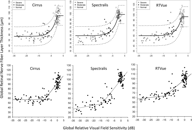
Purpose: To estimate visual field (VF) sensitivity at which retinal nerve fiber layer (RNFL) thinning reaches the measurement floor and at which RNFL stops thinning (change points), the dynamic range of RNFL thickness, and the number of steps from normal to RNFL floor among three optical coherence tomography (OCT) devices.
Methods: Glaucomatous patients (n = 58) and healthy subjects (n = 55-60) prospectively underwent VF testing and RNFL thickness measurement with Cirrus, Spectralis, and RTVue. Change points and corresponding RNFL thicknesses were estimated with simple linear regression (SLR) and Bayesian change point (BCP) analyses. The dynamic range and number of steps to RNFL floor were determined.
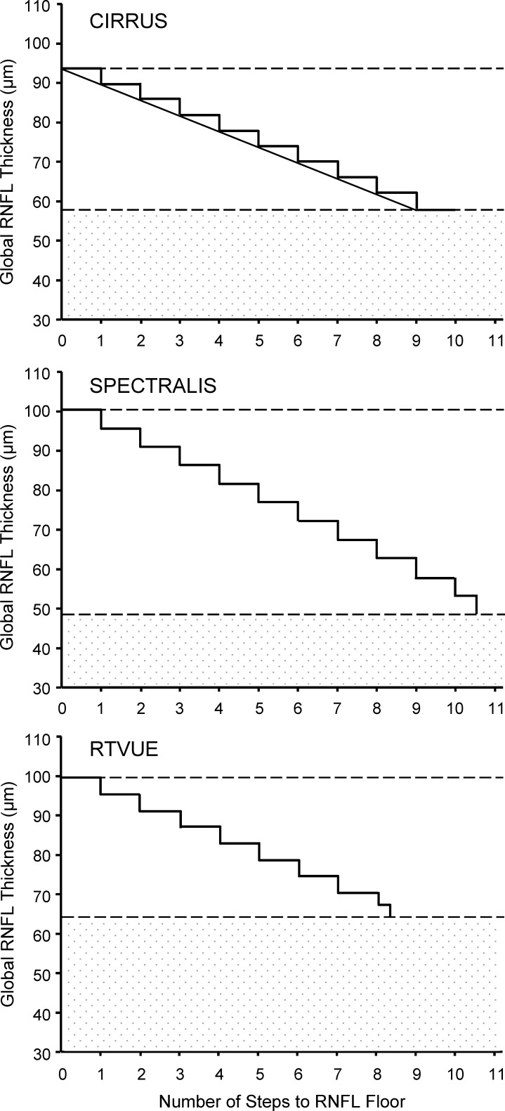
Results: The average VF change points and corresponding residual thickness at the time RNFL stopped thinning were -22.2 dB and 57.0 μm (Cirrus), -25.3 dB and 49.2 μm (Spectralis), and -24.6 dB and 64.7 μm (RTVue). The RNFL dynamic ranges derived from SLR values were wider on Spectralis (52.6 μm) than on Cirrus (35.4 μm) and RTVue (35.5 μm); the corresponding number of steps to reach the RNFL floor were 9.0 on Cirrus, 10.6 on Spectralis, and 8.3 on RTVue.
Conclusions: The relative VF sensitivity at which average RNFL thickness reaches the measurement floor, the residual layer thickness, and RNFL dynamic measurement range differ among the three devices. However, the number of steps from normal to the RNFL thickness floor is comparable.
Figures
References
-
- Garway-Heath DF,, Holder GE,, Fitzke FW,, Hitchings RA. Relationship between electrophysiological psychophysical, and anatomical measurements in glaucoma. Invest Ophthalmol Vis Sci. 2002; 43: 2213–2220. - PubMed
-
- Schlottmann PG,, De Cilla S,, Greenfield DS,, Caprioli J,, Garway-Heath DF. Relationship between visual field sensitivity and retinal nerve fiber layer thickness as measured by scanning laser polarimetry. Invest Ophthalmol Vis Sci. 2004; 45: 1823–1829. - PubMed
-
- Leung CK,, Chong KK,, Chan WM,, et al. Comparative study of retinal nerve fiber layer measurement by StratusOCT and GDx VCC, II: structure/function regression analysis in glaucoma. Invest Ophthalmol Vis Sci. 2005; 46: 3702–3711. - PubMed
Publication types
MeSH terms
Grants and funding
LinkOut - more resources
Full Text Sources
Other Literature Sources
Miscellaneous
