. 2015 Oct 5;4(10):e169.
doi: 10.1038/oncsis.2015.28.

PIK3CA mutations can initiate pancreatic tumorigenesis and are targetable with PI3K inhibitors

Affiliations

PIK3CA mutations can initiate pancreatic tumorigenesis and are targetable with PI3K inhibitors

S N Payne et al. Oncogenesis. .

Abstract

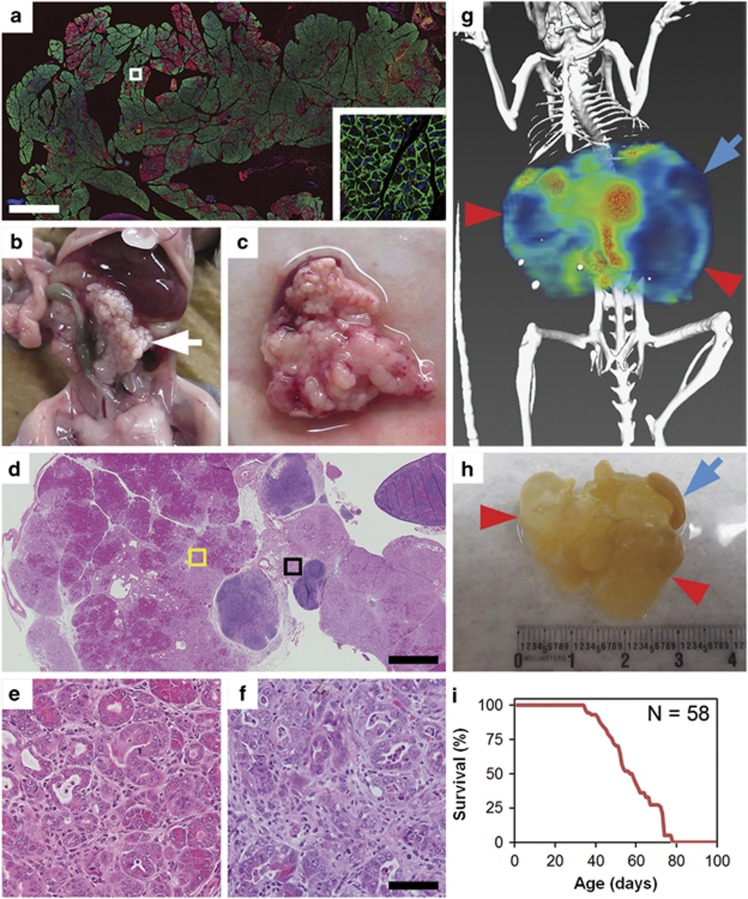
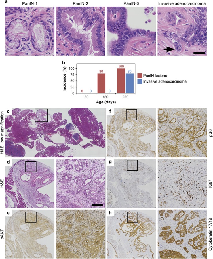
Aberrations in the phosphoinositide 3-kinase (PI3K) signaling pathway have a key role in the pathogenesis of numerous cancers by altering cell growth, metabolism, proliferation and apoptosis. Interest in targeting the PI3K signaling cascade continues, as new agents are being clinically evaluated. PIK3CA mutations result in a constitutively active PI3K and are present in a subset of pancreatic cancers. Here we examine mutant PIK3CA-mediated pancreatic tumorigenesis and the response of PIK3CA mutant pancreatic cancers to dual PI3K/mammalian target of rapamycin (mTOR) inhibition. Two murine models were generated expressing a constitutively active PI3K within the pancreas. An increase in acinar-to-ductal metaplasia and pancreatic intraepithelial neoplasms (PanINs) was identified. In one model these lesions were detected as early as 10 days of age. Invasive pancreatic ductal adenocarcinoma developed in these mice as early as 20 days of age. These cancers were highly sensitive to treatment with dual PI3K/mTOR inhibition. In the second model, PanINs and invasive cancer develop with a greater latency owing to a lesser degree of PI3K pathway activation in this murine model. In addition to PI3K pathway activation, increased ERK1/2 signaling is common in human pancreatic cancers. Phosphorylation of ERK1/2 was also investigated in these models. Phosphorylation of ERK1/2 is demonstrated in the pre-neoplastic lesions and invasive cancers. This activation of ERK1/2 is diminished with dual PI3K/mTOR inhibition. In summary, PIK3CA mutations can initiate pancreatic tumorigenesis and these cancers are particularly sensitive to dual PI3K/mTOR inhibition. Future studies of PI3K pathway inhibitors for patients with PIK3CA mutant pancreatic cancers are warranted.


Figures

Figure 1
Figure 1
The expression of a constitutively active PI3K resulting in pancreatic tumor formation. (a) Pc1 mT/mG1 mice demonstrated the expression of GFP throughout the pancreas, indicating expression of the Cre recombinase at the site of interest. Size bar, 1 mm. Area outlined is shown enlarged × 20 in inset. Pc1 Pik3cap110* mice were allowed to age until moribund; at necropsy large pancreatic tumors were identified: (b) in vivo; (c) whole mount. (d) Histological analysis demonstrated heterogenous masses comprised of dysplatic acinar cells, neoplastic changes, abundant stromal infiltration and immune infiltrates. Size bar, 1mm. Area outlined in yellow is enlarged × 20 in e; the area outlined in black is enlarged × 20 in f. At higher magnification, acinar-to-ductal metaplasia could be identified (e) in addition to invasive pancreatic ductal adenocarcinoma (f). Size bar for e and f, 50 μm. (g and h) Dual hybrid 18F-FDG microPET/CT imaging was used to better understand the complex heterogeneity within these tumors including areas of dense fibrosis with invasive cancer (orange) and areas of cystic dilation (red arrows). Blue arrows denote the spleen. (i) All Pc1 Pik3cap110* mice developed tumors and became moribund at a median age of 53 days.
Figure 2
Figure 2
p110* results in increased activation of the PI3K pathway in the pancreas of Pc1 Pik3cap110* mice compared with age-matched controls. (a) A marked difference in pancreatic morphology is noted with the development of invasive pancreatic cancer surrounded by a dense fibrous infiltrate. The areas of neoplasia had increased phosphorylation of AKT and RPS6 on IHC. An increase in cellular proliferation was also observed based on an increase in nuclear Ki67 staining in the neoplastic cells. Size bar for the top panels, 1 mm. Each lower panel is a × 10 enlargement of the outlined area in the top panel in its respective column or equivalent area in the same sample. (b) The activation of the PI3K pathway in the pancreatic tissue of moribund Pc1 Pik3cap110* mice was also confirmed with immunoblotting. (c) Statistically significant increases in phosphorylation of AKT and RPS6 were identified (P=0.02 and P=0.03, respectively, two-sided Wilcoxon Rank Sum test). Values were normalized to GAPDH.
Figure 3
Figure 3
Pc1 Pik3cap110* mice develop metaplasia, PanINs and invasive cancers at very early ages. PanIN-1 (a), PanIN-2 (b), PanIN-3 (c) and invasive cancers (d) were identified commonly in Pc1 Pik3cap110* mice. Size bar for ad, 50 μm. (e and f) At only 10 days of age cells undergoing acinar-to-ductal metaplasia were observed. (g and h) The transition to a ductal phenotype was confirmed as these cells stained positive for cytokeratin 17/19. Size bar for eh, 50 μm, (i) A time course was performed starting at 10 days of age. At this time point metasplastic lesions were seen throughout the pancreas. These lesions had strong activation of the PI3K cascade as determined by increased phosphorylation of AKT and RPS6. At just 20 days of age invasive ductal adenocarcinoma was identified. These invasive lesions also had increased phsophorylation of AKT and RPS6. Size bars, 500 μm. Each high-magnification images is a × 10 enlargement of the outlined area in the low-magnification image to its immediate left.
Figure 4
Figure 4
Pc1 Pik3caH1047R mice carry a conditional transgene encoding for the Pik3ca H1047R hotspot mutation. This mutation results in a constitutively active PI3K similar to that seen in the Pc1 Pik3cap110*. (a) PanIN lesions were seen in this model in addition to invasive pancreatic ductal adenocarcinoma (arrow). Size bar, 20 μm. (b) This was seen with a greater latency than that observed in the Pc1 Pik3cap110* model with PanIN lesions identified at 150 days of age and most mice revealing invasive cancer by 250 days of age. (c) Abundant fibrous stroma was also seen. Activation of the PI3K signaling cascade was observed in these tumors, including phosphorylation of AKT (e) and RPS6 (f). Increased cellular proliferation was noted on Ki67 staining (g). The transition to ductal cells was confirmed with cytokeratin 17/19 staining (h). (dh) Each medium-resolution photo is a × 4 enlargement of the outlined area in panel c (or equivalent area from the same sample); each high-resolution photo is a × 5 enlargement of the outlined area to its immediate left Size bar for high-resolution panels, 100 μm.
Figure 5
Figure 5
Pancreatic cancers initiated secondary to a constitutively active PI3K are profoundly sensitive to dual PI3K/mTOR inhibition. The pancreatic cancers in Pc1 Pik3cap110* mice are composed of malignant epithelial cells and dense stroma. In untreated mice, phosphorylation of AKT is observed indicating activation of the PI3K pathway (left columns). In addition, these tumors are highly proliferative with an increase in nuclear Ki67 staining (left columns). A cohort of Pc1 Pik3cap110* mice were treated with NVP-BEZ235 by oral gavage. At 6 h post treatment, a decrease in phosphorylation of AKT was identified (middle columns). After 4 days of treatment necropsy was performed and sections of the remaining pancreatic tumor were stained with H&E (right columns). A marked treatment response was observed. There was a significant decrease in the epithelial component leaving the fibrous stroma composing the majority of the remaining tumor. This was also associated with a decrease in phospho AKT and an impressive reduction in Ki67 staining. Size bar, 1mm. Each high-magnification image is a × 5 enlargement of the outlined area to its immediate left.
Figure 6
Figure 6
Activation of the RAS/RAF/MEK/ERK signaling cascade is a hallmark of pancreatic ductal adenocarcinoma and is present in cancers initiated by PI3K. The phosphorylation of ERK1/2 in pancreatic ductal adenocarcinoma is most commonly secondary to activating KRAS mutations. (a) The pancreatic cancers in Pc1 Pik3cap110* mice possess significant phosphorylation of ERK1/2. (b) Metaplastic pancreatic lesions in mice at 10 days of age and the invasive cancers in mice at only 20 days of age also demonstrate activation of ERK1/2. (c) The levels of phosphorylated ERK1/2 are reduced markedly 6 h after treatment of these cancers with NVP-BEZ235. (d) This suppression is maintained following 4 days of treatment. Size bars, 1 mm. Each high-magnification image is a × 10 enlargement of the outlined area to its immediate left.

References

    1. 1Cancer Facts & Figures 2014. American Cancer Society: Atlanta, 2014.
    1. 2Conroy T, Desseigne F, Ychou M, Bouche O, Guimbaud R, Becouarn Y et al. FOLFIRINOX versus gemcitabine for metastatic pancreatic cancer. N Engl J Med 2011; 364: 1817–1825. - PubMed
    1. 3Von Hoff DD, Ervin T, Arena FP, Chiorean EG, Infante J, Moore M et al. Increased survival in pancreatic cancer with nab-paclitaxel plus gemcitabine. N Engl J Med 2013; 369: 1691–1703. - PMC - PubMed
    1. 4Teague A, Lim KH, Wang-Gillam A. Advanced pancreatic adenocarcinoma: a review of current treatment strategies and developing therapies. Ther Adv Med Oncol 2015; 7: 68–84. - PMC - PubMed
    1. 5Cataldo VD, Gibbons DL, Perez-Soler R, Quintas-Cardama A. Treatment of non-small-cell lung cancer with erlotinib or gefitinib. N Engl J Med 2011; 364: 947–955. - PubMed