. 2016 Jan;29(1):68-80.
doi: 10.1111/pcmr.12426. Epub 2015 Nov 3.

Effective intra-S checkpoint responses to UVC in primary human melanocytes and melanoma cell lines

Affiliations

Effective intra-S checkpoint responses to UVC in primary human melanocytes and melanoma cell lines

Marila Cordeiro-Stone et al. Pigment Cell Melanoma Res. 2016 Jan.

Abstract

The objective of this study was to assess potential functional attenuation or inactivation of the intra-S checkpoint during melanoma development. Proliferating cultures of skin melanocytes, fibroblasts, and melanoma cell lines were exposed to increasing fluences of UVC and intra-S checkpoint responses were quantified. Melanocytes displayed stereotypic intra-S checkpoint responses to UVC qualitatively and quantitatively equivalent to those previously demonstrated in skin fibroblasts. In comparison with fibroblasts, primary melanocytes displayed reduced UVC-induced inhibition of DNA strand growth and enhanced degradation of p21Waf1 after UVC, suggestive of enhanced bypass of UVC-induced DNA photoproducts. All nine melanoma cell lines examined, including those with activating mutations in BRAF or NRAS oncogenes, also displayed proficiency in activation of the intra-S checkpoint in response to UVC irradiation. The results indicate that bypass of oncogene-induced senescence during melanoma development was not associated with inactivation of the intra-S checkpoint response to UVC-induced DNA replication stress.

Keywords: DNA replication; human; intra-S checkpoint; melanocyte; melanoma; replicon initiation; ultraviolet radiation.


Figures

Figure 1
Figure 1
Velocity sedimentation analysis discloses both active and passive responses to UVC-induced DNA damage. Normal human melanocytes with nuclear DNA homogeneously labeled with [14C]thymidine were exposed to increasing fluences of UVC, incubated for 30 min, pulse-labeled for 15 min with [3H]thymidine, and lysed on top of 5–20% alkaline sucrose gradients. After sedimentation and fractionation, acid-insoluble radioactivity was quantified. The graphs illustrate the size distributions of radiolabeled single-stranded DNA in NHM16 (A), NHM18 (B) and NHM28 (C) melanocyte cultures exposed to zero (sham; white-filled circles), 1 J/m2 (black-filled triangles), or 10 J/m2 (gray-filled squares) of 254 nm UVC radiation (see Supplementary Figure S2 for results with other UVC fluences). Normalized 3H-CPM represents the radioactivity incorporated into nascent DNA of various sizes during the 15-min pulse with [3H]thymidine, relative to the total number of cells added to each gradient (as measured by the total 14C-CPM) and to the highest value in the experimental group. This normalization also illustrates the extent of DNA synthesis inhibition by each of the UVC fluences. The profile observed in the cultures exposed to 1 J/m2 UVC, compared to the sham control, revealed (as illustrated in A and B) a selective inhibition in molecules with sizes less than 60 kb (marked by the arrow), consistent with the intra-S checkpoint response of inhibition of replicon initiation (see also Fig. 2A). A larger and cytotoxic fluence of UVC (10 J/m2) produced a strong inhibition of DNA synthesis in active replicons (gradient fractions 5–17), due to blockage of DNA polymerases at template lesions (passive inhibition) and active checkpoint signaling, with a concomitant appearance of a peak of abnormally small DNA (in fractions 21–25) that heralds the presence of daughter-strand gaps. The smaller size of nascent DNA seen in NHM28 may be the result of enhanced fluorescent-light-induced fragmentation of DNA during cell lysis due to high melanin content of the cells.
Figure 2
Figure 2
UVC-induced inhibition of DNA strand growth in diploid human cells. (A) Results of a second velocity sedimentation experiment with NHM28 using higher fluences of UVC (Sham, white-filled circles; 3.0 J/m2, black-filled diamonds; 5 J/m2, black-filled circles; 10 J/m2, gray-filled squares; 15 J/m2, white-filled triangles). Note the evidence for selective inhibition of low molecular weight nascent DNA (fractions 20–24) upon irradiation with 3 J/m2 UVC (inhibition of replicon initiation). As the incident UVC fluence was increased, radiolabeled precursor incorporation in large molecular weight DNA intermediates (range between the two arrows) decreased. The equivalent ranges of fractions from all gradients with the same cell type were used to quantify the UVC-dependent inhibition of DNA strand growth. (B) The decrease in radiolabel incorporation in high molecular weight intermediates of DNA replication, relative to the non-irradiated (sham) controls (average of duplicate experiments) was plotted against UVC fluence. Results with normal melanocytes (filled symbols: gray circle, NHM16; gray diamond, NHM18; and black triangles, NHM28) were compared to those previously published for two NHF lines (squares) and an XP-V cell line (circles) (King et al., 2005). The slopes determined for the NHF and XP-V lines were equivalent to slopes quantified in different NHF and XP-V strains in an earlier study (Kaufmann and Cleaver, 1981).
Figure 3
Figure 3
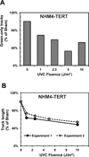
UVC-induced inhibition of replicon initiation and DNA chain elongation in an immortalized normal human melanocyte line. NHM4-hTERT in log phase growth were pulse labeled with IdU for 15 min, exposed to the indicated fluences of UVC, then incubated in CldU-containing medium for 30 min. Individual DNA fibers were spread and immuno-stained as previously described (Chastain et al., 2006). Replication tracts containing IdU (pre-label) were stained red and those containing CldU (post-label) were stained green. (A) Frequency of green-only tracks (replicons initiated after UVC exposure) in irradiated samples, relative to the sham control. The reduction in this class of labeled fibers indicates the degree of inhibition of replicon initiation by UVC. Results represent the average of two independent experiments in which 345 to 1036 individual fibers were counted in each sample. (B) UVC-dependent inhibition of DNA chain elongation. Labeled fibers with a green portion (CldU label) attached to a red portion (IdU) indicated replication units that were active before UVC exposure and continued to synthesize DNA after irradiation (green portion). The average length of the green portion of red-green tracks was measured in 39–71 individual fibers in each sample of two independent experiments. The inhibition curves show two distinct components: the sharp decrease in track length after 1 J/m2 UVC appears to reflect the intra-S checkpoint-dependent, active slowing of DNA chain elongation; as the UVC fluence was increased further, the subsequent reduction in relative track length seems to include the contribution of passive inhibition of chain elongation at DNA template lesions.
Figure 4
Figure 4
CPD dosimetry in melanocytes and fibroblasts. Monolayer cultures of NHF1 fibroblasts (white squares), NHM16 melanocytes (gray-filled circles) and NHM28 melanocytes (black-filled triangles) were irradiated with the indicated fluences of UVC, and harvested immediately afterwards for DNA purification and CPD quantification by immunoblotting (Sproul et al., 2014). Illustrated results represent the averages of two independent experiments for each of the identified cultured cells. Quantification of melanin content (Watts et al., 1981) yielded the following values: NHM16, 15 µg/106 cells; NHM28, 39 µg/106 cells.
Figure 5
Figure 5
DNA damage response markers in normal fibroblasts and melanocytes. Whole cell extracts were prepared from NHM16 and NHM18 melanocytes 45 min after exposure to the indicated fluences of UVC. Parallel experiments were also done with NHF1-hTERT and NHF10-hTERT fibroblasts. After gel electrophoresis and transfer to nitrocellulose, the membranes were probed with the indicated antibodies (see METHODS for details). Phospho-proteins were probed before the same membranes were stripped and then re-probed with the antibodies recognizing the modified and unmodified forms of the same proteins. Short (s) and long (l) exposures were used to detect the P-ATM signals. Image J was used to quantify the immunoblot signals from unsaturated X-ray films and the ratios of the phosphorylated form over the total for each protein were determined and expressed relative to the same parameter determined in the sham-treated control (set as 1.0).
Figure 6
Figure 6
Velocity sedimentation analysis of DNA replication in melanoma cell lines. Three cell lines were chosen (without previous knowledge of their response to UVC at the level of DNA replication) from each of three melanoma groups, based on the mutation status at the BRAF and NRAS loci: (A) Expressing wild type B-Raf and N-Ras; (B) Expressing a mutant N-Ras; (C) Expressing a mutant B-Raf. The experimental procedure was the same as described in the legend to Figure 1. The illustrated profiles are those for the sham-treated sample (white circles), and for the cells irradiated with 1 J m2 UVC (black-filled triangles) or 10 J/m2 UVC (gray-filled squares). Supplementary Figure S4 includes the results with 2.5 and 5 J/m2 UVC. Note that after 1 J/m2 UVC, all cell lines inhibited incorporation into nascent DNA of low molecular weight (inhibition of replicon initiation), albeit to different degrees. The pattern seen with RPMI 8322 was distinct from the other cell lines in that a minor peak of abnormally small DNA was already apparent at 1 J/m2 and when the UVC fluence was increased to 10 J/m2 the peak of abnormally small DNA rose above the control profile. SKMel-23 was first scored as wild-type for BRAF and NRAS. After subsequent studies revealed behavior similar to B-Raf-mutant lines (Sambade et al., 2011), reanalysis identified a rare BRAF mutation (G466A).
Figure 7
Figure 7
UVC induction of Chk1 phosphorylation in melanoma cell lines. The experimental details were as described in the legend to Figure 5.
Figure 8
Figure 8
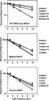
Inhibition of DNA strand growth by UVC in melanoma cell lines. The average results of inhibition of incorporation of radiolabeled [3H]thymidine in high molecular weight nascent DNA species (see Figure 2) from 2–3 independent experiments for each cell line were plotted against UVC fluences (no significant differences in CPD densities were expected among the different melanomas and NHM16 and NHM18 – see text). The slopes determined for each line are reported in Table 1. Only RPMI 8322 displayed results that were clearly distinct from the normal melanocytes and the other melanoma cell lines (p<0.0001).
Figure 9
Figure 9
Relative abundance of DNA polymerase eta. Chromatin was isolated from the indicated cultured cells 45 min after sham treatment (−) or irradiation with 10 J/m2 UVC (+). Twenty five micrograms of chromatin protein from each sample were loaded onto polyacrylamide gels. Following gel electrophoresis and transfer to a nitrocellulose membrane, the chromatin-associated proteins were probed with antibodies against DNA pol eta, PCNA and α-tubulin. XP-V fibroblasts were included as a negative control for DNA pol eta expression. Pixel intensity values in unsaturated X-ray films were quantified for each protein. The levels of DNA pol eta were expressed relative to PCNA and α-tubulin.

References

    1. Abbas T, Sivaprasad U, Terai K, Amador V, Pagano M, Dutta A. PCNA-dependent regulation of p21 ubiquitylation and degradation via the CRL4Cdt2 ubiquitin ligase complex. Genes & development. 2008;22:2496–2506. - PMC - PubMed
    1. Bower JJ, Zhou Y, Zhou T, Simpson DA, Arlander SJ, Paules RS, Cordeiro-Stone M, Kaufmann WK. Revised genetic requirements for the decatenation G2 checkpoint: the role of ATM. Cell cycle. 2010;9:1617–1628. - PMC - PubMed
    1. Brown EJ, Baltimore D. ATR disruption leads to chromosomal fragmentation and early embryonic lethality. Genes & development. 2000;14:397–402. - PMC - PubMed
    1. Carson C, Omolo B, Chu H, Zhou Y, Sambade MJ, Peters EC, Tompkins P, Simpson DA, Thomas NE, Fan C, et al. A prognostic signature of defective p53-dependent G1 checkpoint function in melanoma cell lines. Pigment cell & melanoma research. 2012;25:514–526. - PMC - PubMed
    1. Chastain PD, Brylawski BP, 2nd, Zhou YC, Rao S, Chu H, Ibrahim JG, Kaufmann WK, Cordeiro-Stone M. DNA damage checkpoint responses in the S phase of synchronized diploid human fibroblasts. Photochemistry and photobiology. 2015;91:109–116. - PMC - PubMed

Publication types

MeSH terms