. 2015 Oct 5;10(10):e0139418.
doi: 10.1371/journal.pone.0139418. eCollection 2015.

The SET Domain Is Essential for Metnase Functions in Replication Restart and the 5' End of SS-Overhang Cleavage

Affiliations

The SET Domain Is Essential for Metnase Functions in Replication Restart and the 5' End of SS-Overhang Cleavage

Hyun-Suk Kim et al. PLoS One. .

Abstract

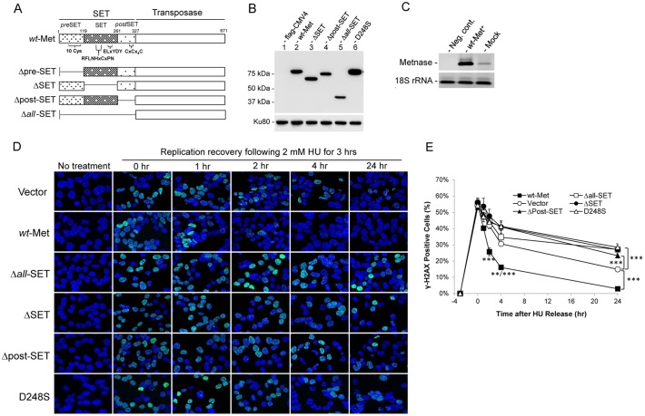
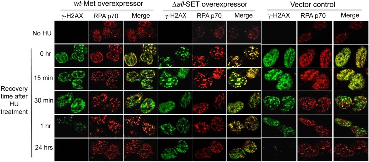
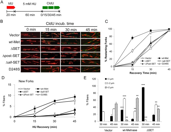
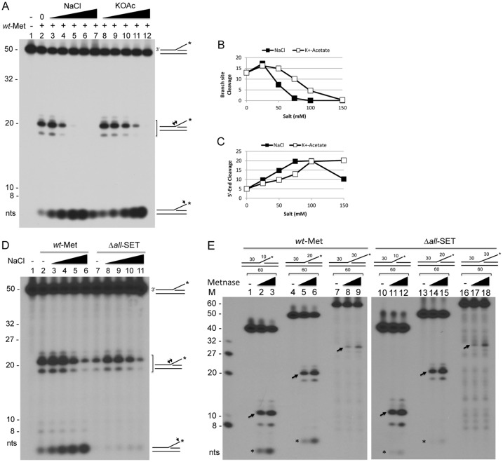
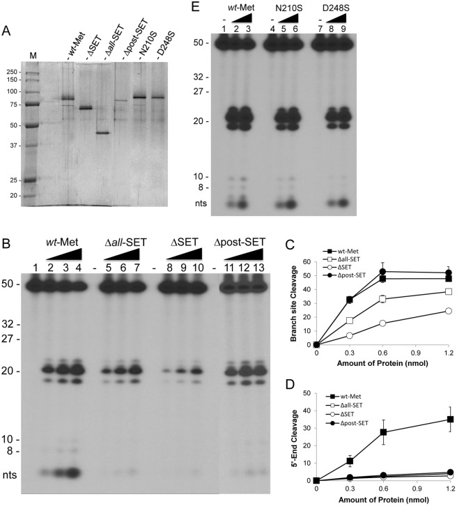
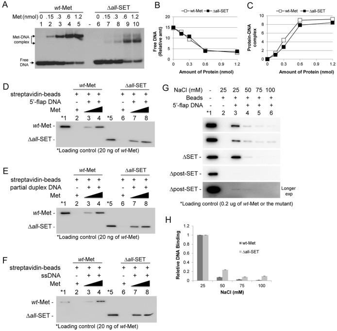
Metnase (also known as SETMAR) is a chimeric SET-transposase protein that plays essential role(s) in non-homologous end joining (NHEJ) repair and replication fork restart. Although the SET domain possesses histone H3 lysine 36 dimethylation (H3K36me2) activity associated with an improved association of early repair components for NHEJ, its role in replication restart is less clear. Here we show that the SET domain is necessary for the recovery from DNA damage at the replication forks following hydroxyurea (HU) treatment. Cells overexpressing the SET deletion mutant caused a delay in fork restart after HU release. Our In vitro study revealed that the SET domain but not the H3K36me2 activity is required for the 5' end of ss-overhang cleavage with fork and non-fork DNA without affecting the Metnase-DNA interaction. Together, our results suggest that the Metnase SET domain has a positive role in restart of replication fork and the 5' end of ss-overhang cleavage, providing a new insight into the functional interaction of the SET and the transposase domains.


Conflict of interest statement

Competing Interests: The authors have declared that no competing interests exist.

Figures

Fig 1
Fig 1. The Metnase SET domain is necessary for replication recovery after long exposure to hydroxyurea (HU).
(A) Schematic diagram of Metnase. The Metnase SET domain comprises pre-SET (aa 14–118), SET (aa 119–260), and post-SET (aa 261–326) domains. The pre SET domain contains a cysteine-rich putative Zn+2 binding motif (10 Cys), while the SET domain has the SAM binding motif (207-RFLNHxCxPN----ELxYDY-248) for histone lysine methyltransferase activity. The post-SET domain also contains a cysteine-rich putative Zn+2 binding motif (CxCx4C). (B) Western blot analysis of flag-tagged wt-Metnase and the SET mutants that were stably expressed in HEK293 cells. Thirty μg of cell extracts were loaded onto 10% SDS-PAGE for immunoblot analysis. Ku80 was used as a loading control. (C) Metnase expression in control HEK293 cells (mock) and wt-Metnase overexpressor (wt-Met+) was analyzed by RT-PCR. (D) Representative confocal microscope images of HEK293 cells stably transfected with pCMV4 vector (top row), wt-Metnase (second row), and the SET mutants (third to sixth row) following HU treatment. After treatment of 2 mM HU for 3 hrs, cells were released into fresh media at indicated times, stained with DAPI (blue) and an antibody to γ-H2AX (green), and imaged by confocal microscopy. (E) Quantitation of γ-H2AX-positive cells in panel D. Plots were average percentages (±SD) of γ-H2AX-positive cells. An average of 200 cells were counted per slide, 6 slides per experiment. **, P<0.01; ***, P<0.005, t tests.
Fig 2
Fig 2. The Metnase SET domain is necessary for Metnase function in damage recovery prior to replication restart following HU treatment.
Representative images of γ-H2AX and RPA p70 foci in HEK293 cells stably transfected with pCMV4 vector, wt-Metnase, and the SET deletion mutant (Δall-SET), as indicated on the top. After treatment with 2 mM HU for 3 hrs, cells were released into fresh media at indicated times, stained with DAPI (blue) and antibodies to γ-H2AX (green) and RPA p70 (red), and imaged by confocal microscopy.
Fig 3
Fig 3. The Metnase SET domain has a crucial role in replication fork restart following HU treatment.
(A) Dual labeling protocol for replication restart using DNA fiber analysis. Cells were pulse-labeled with IdU (red) for 20 min, treated with 5 mM HU for 60 min, and labeled with CldU (green) for 0, 15, 30, and 45 min. (B) Representative confocal microscope images of replication tracks from HEK293 cells stably expressing vector (top row), wt-Metnase (2nd row), and the SET deletion/substitution mutants (3rd- 6th rows). Cells were treated with 5 mM HU prior to pulse labeling with CldU for indicated amounts of time. (C-D) Average percentage (±SD) of restarted forks (red plus green; panel C) and new forks (green only; panel D) for three independent experiments in which 150–200 fibers were scored per each determination. (E) Fiber lengths were measured by using Olympus FV1—ASW 3.0 software. Average lengths of 150–200 fibers were scored in triplicates per each determination. **, P<0.01; ***, P<0.005 (compared with vector control), t tests.
Fig 4
Fig 4. The Metnase SET domain is necessary for the 5’-end cleavage of ss-overhang DNA.
(A) Silver staining of purified wt-Metnase (wt-MET), the SET deletion mutant (Δall-SET), and a nuclease-dead mutant (D483A) following 10% SDS-PAGE. (B) The SET domain is essential for cleavage of the 5’-flap DNA. Reaction mixtures (20 μl) containing the 5’-32P-labeled flap DNA (60 fmol) and increasing amounts of wt-Metnase or the mutant (Δall-SET or D483A) were incubated at 37°C in the presence of 2 mM MgCl2 for 90 min, and cleavage products were analyzed by 12% PAGE containing 8M urea. Numbers on the left indicated DNA size makers. (C-D) Cleavage of the branch site (panel C) and the 5’ end of ss-overhang (panel D) of a 5’-flap DNA shown in Panel B was quantified using a PhosphorImager and ImageQuant software (Molecular Dynamics). (E) Cleavage of the 5’ end of the 5’-32P-labeled partial duplex DNA with wt-Metnase and the SET deletion mutant. Indicated amount of wt-Metnase or the Δall-SET was incubated with the 5’-32P-labeled (*) 5’-ss-overhang DNA (60 fmol) for 90 min at 37°C prior to 12% PAGE analysis (+ 8M urea). Arrows on the right mark the cleavage sites on the 5’-32P-labeled (*) DNA.
Fig 5
Fig 5. The Metnase SET domain is required for a preferential cleavage of the 5’ end of a 5’-flap DNA in the presence of high salt.
(A) Metnase prefers cleavage of the branch site at low salt, while it likes cleavage of the 5’-end of a 5’-flap DNA in the presence of high salt. Wt-Metnase (50 ng) was incubated with 60 fmol of the 5’-32P-labeled (*) DNA for 120 min in the presence of increasing concentrations (0, 25, 50, 75, 100, and 150 mM) of NaCl or potassium acetate (KOAc) prior to 12% denatured PAGE (+ 8M urea) analysis. Arrows on the right mark the cleavage sites on the 32P-labeled (*) 5’-flap DNA. (B-C) Metnase-mediated cleavage of the branch site (panel B) and the 5’ end of ss-overhang (panel C) of a 5’-flap DNA shown in Panel A was quantified. (D) Cleavage of the 5’ end of a 5’-32P-flap DNA with wt-Metnase and the SET deletion mutant in the presence of varying salt concentrations. Fifty ng of wt-Metnase or the Δall-SET was incubated with a 5’-32P-flap DNA in the presence of increasing concentrations (0, 25, 50, 75, and 100 mM) of NaCl. Arrows on the right mark the cleavage sites on the 5’-32P-labeled (*) flap DNA. (E) Metnase exhibits branch site cleavage of the 5’-flap DNA with different sizes of ss-overhang. Wt-Metnase (50 or 100 ng; lanes 1–9) or the SET deletion mutant (lanes 10–18) was incubated with the 5’-32P-labeled 5’-flap DNA in different length of ss-overhang (10-mer, lanes 1–3 & 10–12; 20-mer, lanes 4–6 & 13–15; 30-mer, lanes 7–9 & 16–18). Arrows mark the major branch site cleavage with the 32P-labeled 5’-flap DNA. M on the left represents DNA markers.
Fig 6
Fig 6. The Metnase SET domain but not its HLMT activity is essential for cleavage of the 5’ end of a 5’-flap DNA.
(A) Silver staining of purified wt-Metnase (wt-MET) and the SET deletion and the substitution mutants following 10% SDS-PAGE. (B) Cleavage of the 5’ end of a 5’-flap DNA with the SET deletion and the substitution mutants. Increasing amounts of wt-Metnase and the SET domain deletion mutant (panel B) were incubated with 60 fmol of a 5’-32P-flap DNA for 120 min prior to 12% denatured PAGE (+ 8 M urea) analysis. (C-D) Metnase-mediated cleavage of the branch site (panel C) and the 5’ end of ss-overhang (panel D) of a 5’-flap DNA shown in Panel B was quantified. (E) Increasing amounts of wt-Metnase and the substitution mutants lacking HLMT activity (N210S & D248S) were examined for cleavage of the 5’-flap DNA.
Fig 7
Fig 7. The Metnase SET domain is not involved in the Metnase-DNA interaction.
(A) Indicated amounts of wt-Metnase or the Δall-SET mutant were incubated with 400 fmol of 5’-32P-labeled TIR DNA. Following 15 min incubation at 25°C, the protein–DNA complexes were analyzed by 5% native PAGE in the presence of 1X TBE. (B-C) Quantitation of free DNA and the Metnase-TIR complex. For quantitation, individual bands were excised from dried gel and measured for radioactivity. (D-F) Interaction of wt-Metnase and the SET deletion mutant (Δall-SET) with DNA using Streptavidine pulldown assay. Flag-tagged wt-Metnase or Δall-SET protein (1.0 and 2.0 μg) was incubated with 50 pmol of the 3’-biotinylated 5’-flap DNA (panel D), a partial duplex DNA (panel E), or ssDNA (panel F) for protein-DNA binding by Streptavidin-agarose beads (see Experimental Procedures for the details). The protein-DNA interaction was analyzed by Western blot using an anti-flag antibody. (G) Salt sensitivity of wt-Metnase and the SET deletion mutants in their interaction with a 5’-flap DNA. Wt-Metnase or the SET deletion mutant (2.0 μg) was incubated with 50 pmol of the 3’-biotinylated 5’-flap DNA in the presence of varying concentrations of NaCl prior to DNA pull-down with streptavidin-agarose beads. The protein-DNA interaction was analyzed by Western blot using an anti-flag antibody. (H) The protein-DNA complexes (panel G) were quantified by Molecular Imager ChemiDoc XRS using Quantity One® analysis software program (BioRad).

Similar articles

Cited by

References

    1. Beck BD, Lee SS, Wiliamson E, Hromas RA, Lee SH. Biochemical Characterization of Metnase's Endonuclease Activity and Its Role in NHEJ Repair. Biochemistry. 2011;50(20):4360–70. 10.1021/Bi200333k ISI:000290594000015. - DOI - PMC - PubMed
    1. De Haro LP, Wray J, Williamson EA, Durant ST, Corwin L, Gentry AC, et al. Metnase promotes restart and repair of stalled and collapsed replication forks. Nucleic acids research. 2010;38(17):5681–91. 10.1093/nar/gkq339 - DOI - PMC - PubMed
    1. Fnu S, Williamson EA, De Haro LP, Brenneman M, Wray J, Shaheen M, et al. Methylation of histone H3 lysine 36 enhances DNA repair by nonhomologous end-joining. Proceedings of the National Academy of Sciences of the United States of America. 2011;108(2):540–5. Epub 2010/12/29. 10.1073/pnas.1013571108 - DOI - PMC - PubMed
    1. Hromas R, Wray J, Lee SH, Martinez L, Farrington J, Corwin LK, et al. The human set and transposase domain protein Metnase interacts with DNA Ligase IV and enhances the efficiency and accuracy of non-homologous end-joining. DNA repair. 2008;7(12):1927–37. Epub 2008/09/09. doi: S1568-7864(08)00290-5 [pii]10.1016/j.dnarep.2008.08.002 - DOI - PMC - PubMed
    1. Kim HS, Chen Q, Kim SK, Nickoloff JA, Hromas R, Georgiadis MM, et al. The DDN catalytic motif is required for Metnase functions in non-homologous end joining (NHEJ) repair and replication restart. The Journal of biological chemistry. 2014;289(15):10930–8. 10.1074/jbc.M113.533216 - DOI - PMC - PubMed

Publication types

MeSH terms

LinkOut - more resources