. 2015 Oct 5:6:8357.
doi: 10.1038/ncomms9357.

MAP4K family kinases act in parallel to MST1/2 to activate LATS1/2 in the Hippo pathway

Affiliations

MAP4K family kinases act in parallel to MST1/2 to activate LATS1/2 in the Hippo pathway

Zhipeng Meng et al. Nat Commun. .

Abstract

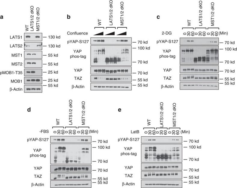
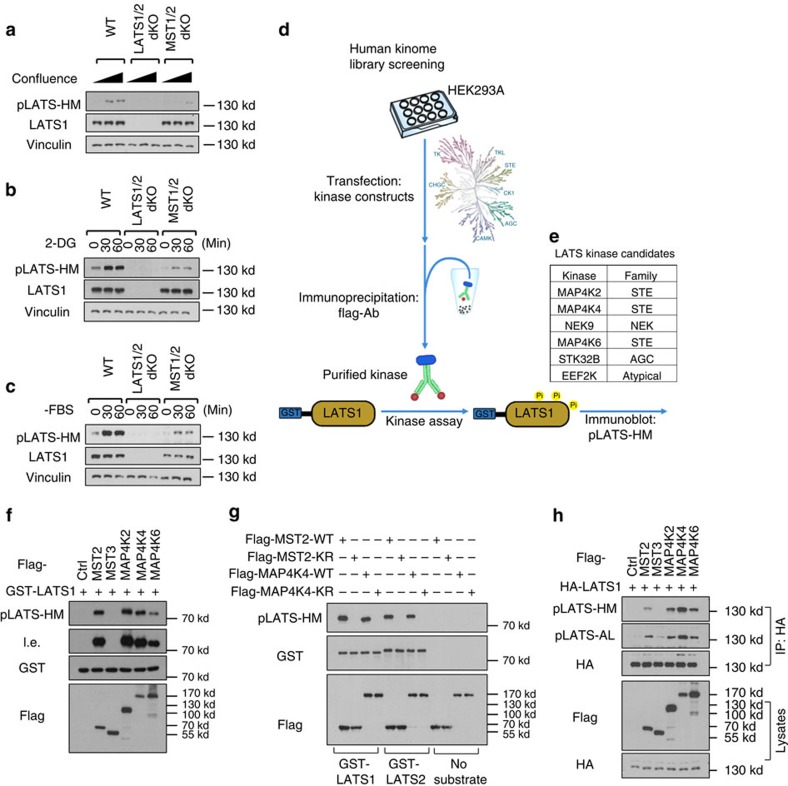
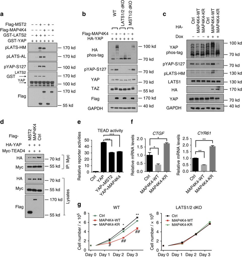
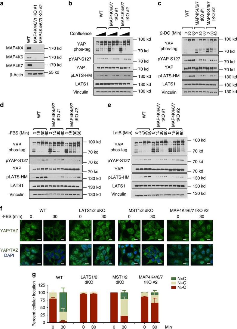
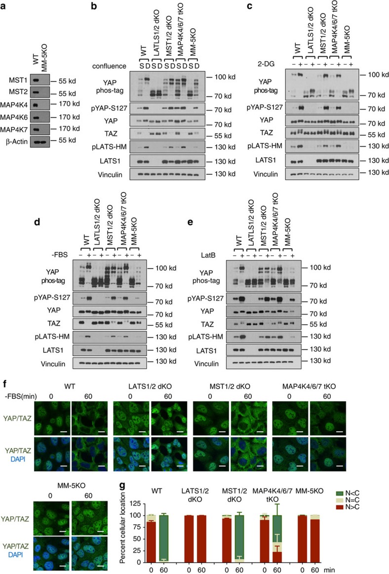
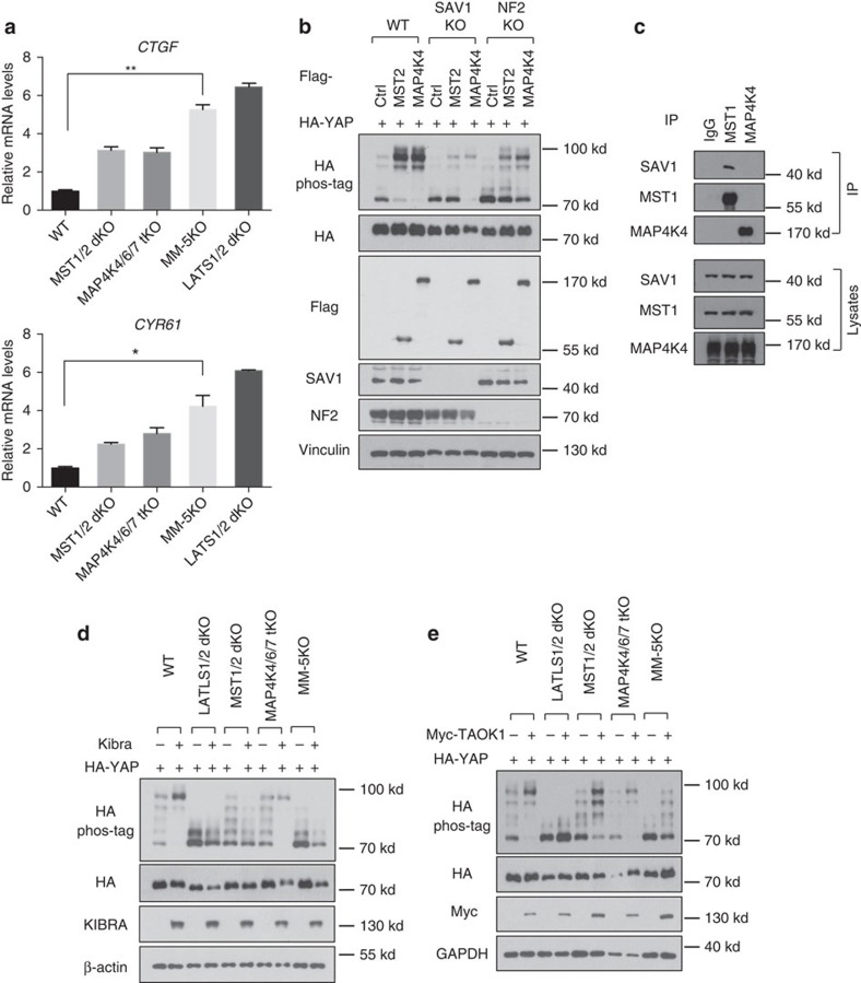
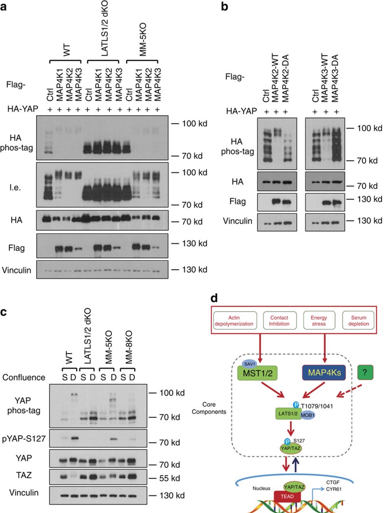
The Hippo pathway plays a central role in tissue homoeostasis, and its dysregulation contributes to tumorigenesis. Core components of the Hippo pathway include a kinase cascade of MST1/2 and LATS1/2 and the transcription co-activators YAP/TAZ. In response to stimulation, LATS1/2 phosphorylate and inhibit YAP/TAZ, the main effectors of the Hippo pathway. Accumulating evidence suggests that MST1/2 are not required for the regulation of YAP/TAZ. Here we show that deletion of LATS1/2 but not MST1/2 abolishes YAP/TAZ phosphorylation. We have identified MAP4K family members--Drosophila Happyhour homologues MAP4K1/2/3 and Misshapen homologues MAP4K4/6/7-as direct LATS1/2-activating kinases. Combined deletion of MAP4Ks and MST1/2, but neither alone, suppresses phosphorylation of LATS1/2 and YAP/TAZ in response to a wide range of signals. Our results demonstrate that MAP4Ks act in parallel to and are partially redundant with MST1/2 in the regulation of LATS1/2 and YAP/TAZ, and establish MAP4Ks as components of the expanded Hippo pathway.


Figures

Figure 1
Figure 1. LATS1/2, but not MST1/2, are essential for YAP regulation by various signals
(a) Immunoblots showing CRISPR-mediated deletion of LATS1/2 and MST1/2 in HEK293A cells. The signal of pMOB1-Threonine 35 (T35) is absent in MST1/2 double knockout (dKO) cells but not in LATS1/2-dKO or wild-type (WT) cells. (b) Contact inhibition induces YAP phosphorylation and TAZ degradation in MST1/2-dKO but not LATS1/2-dKO cells. 1.5 × 10⁵, 6 × 10⁵, and 8 × 10⁵ cells per well were seeded on six-well plates. 24 h later, the cells were collected for immunoblot and phos-tag gel analyses. (c) Energy stress still induces YAP phosphorylation in MST1/2-dKO. Cells under low confluence (1.5 × 10⁵ cells per well were seeded onto six-well plates 24 h before the treatment) were treated with 25 mM 2-DG for 30 or 60 min. (d) Serum depletion induces YAP phosphorylation and TAZ degradation in MST1/2-dKO cells. Cells under low confluence were incubated with serum-free culture medium for 30 or 60 min. (e) YAP phosphorylation is induced by actin depolymerization in the absence of MST1/2. Cells were treated with 0.2 μg ml−1 Latrunculin B (LatB) for 30 or 60 min.
Figure 2
Figure 2. In vitro screen identified MAP4Ks as candidate kinases for LATS1/2.
(a) LATS1/2 phosphorylation is still induced, though weaker, by high cell confluence in MST1/2-dKO cells. An antibody targeting the hydrophobic motif of LATS1/2 kinase domain (pLATS-HM) was used for immunoblotting. (b) Energy stress by 2-DG induces MST1/2-independent LATS1/2 phosphorylation. (c) Serum starvation induces MST1/2-independent LATS1/2 phosphorylation. (d) A scheme of the in vitro human kinome screening for LATS1/2-HM kinases. Flag-tagged individual kinases were expressed and purified from HEK293A cells by anti-flag immunoprecipitation under high stringency, and then applied to the in vitro kinase assays with the truncated form of recombinant human LATS1 as substrates. Phosphorylation of the LATS1-HM was detected with the phospho-specific antibody. (e) A list of kinases that can phosphorylate LATS1-HM based on the in vitro kinome screen. (f) A representative image of the kinase assay is shown. MAP4K2/4/6 phosphorylate the truncated form of recombinant LATS1 at its hydrophobic motif. l.e., long exposure of films. (g) Kinase activity of MAP4K4 is required for its ability to phosphorylate human LATS1 and mouse LATS2. Wild-type and kinase-dead (KR) MST2 and MAP4K4 proteins purified from HEK293A cells were used for the kinase assays. (h) Overexpression of MAP4K2/4/6 induces LATS1 phosphorylation at both its hydrophobic motif (HM) and activation loop (AL) in the HEK293A cells. HA-LATS1 was immunoprecipitated from lysates of the HEK293A cells transfected with the kinase expression plasmids, and then examined for its phosphorylation.
Figure 3
Figure 3. MAP4K4 acts through LATS to phosphorylate and inhibit YAP.
(a) A sequential kinase assay demonstrates that MAP4K4 activates LATS2 by in vitro phosphorylation. The hypophosphorylated and inactive GST-tagged full-length LATS2 purified from the HEK293A cells treated with fresh FBS for 45 min was incubated with immunoprecipitated Flag-MST2 or Flag-MAP4K4 in vitro in the presence of ATP. Recombinant GST-YAP was thereafter added to the reaction to measure GST-LATS2 kinase activity. (b) MAP4K4 overexpression induces YAP phosphorylation in a LATS1/2-dependent manner in HEK293A cells. (c) Doxycycline (Dox) inducible expression shows that the kinase activity of MAP4K4 is required to increase YAP and LATS phosphorylation in HEK293A cells. (d) MAP4K4 reduces YAP-TEAD interaction in HEK293A cells. HA-YAP, Myc-TEAD4, and MAP4K4 or MST2 constructs were co-transfected into HEK293A cells. TEAD4 was thereafter immunoprecipitated with anti-Myc antibody followed by immunoblotting with anti-HA antibody. (e) MAP4K4 suppresses TEAD reporter activity. The constructs expressing the indicated genes were co-transfected with a TEAD firefly luciferase reporter into HEK293A cells. 3 replicates are included in this experiment. The error bars represent s.d. ***P<0.001; student's t-test was applied. (f) MAP4K4 suppresses the expression of YAP target genes CTGF and CYR61. The cells with tetracycline-inducible MAP4K4 overexpression were incubated with doxycycline for 24 h, and then their CTGF and CYR61 expression were measured by quantitative real-time PCRs. Two replicates are included in this experiment. The error bars represent s.d. *P<0.05; **P<0.01; Student's t-test was applied. (g) MAP4K4 inhibits cell growth in a manner dependent on LATS1/2. WT and LATS1/2-dKO HEK293A cells were transfected with MAP4K4 and its KR mutant constructs. The cell number was counted every 24 h. 3 replicates are included in this experiment. The error bars represent s.e.m. *P<0.05; **P<0.01; MAP4K4-KR versus control. #P<0.05; ##P<0.01; MAP4K4-WT vs. control. Student's t-test was applied.
Figure 4
Figure 4. Deletion of MAP4K4/6/7 decreases YAP phosphorylation.
(a) Immunoblot shows the deletion of MAP4K4/6/7 in HEK293A cells. Two independent clones (#1 and #2) are shown. (b) High density-induced LATS and YAP phosphorylation is not altered in MAP4K4/6/7-tKO cells. (c) Energy stress-induced LATS and YAP phosphorylation is slightly reduced in MAP4K4/6/7-tKO cells. (d) Serum starvation-induced LATS and YAP phosphorylation is decreased in MAP4K4/6/7-tKO cells. (e) Actin depolymerization-induced YAP phosphorylation is suppressed by deletion of MAP4K4/6/7 in HEK293A cells. (f) Serum deprivation-induced YAP cytoplasmic translocation is suppressed by MAP4K4/6/7 deletion. Cells were cultured in fresh DMEM containing 10% FBS for 90 min before the serum deprivation. The cells were fixed after 30 min serum depletion and processed for immunofluorescence with anti-YAP/TAZ antibody. Scale bars, 10 μm. (g) Quantification of percentage of the cells with more nuclear (N) or cytosolic YAP/TAZ (C) signals was performed in three randomly chosen fields for each treatment. Typically, each field contains 80–150 cells and are all counted.
Figure 5
Figure 5. Deletion of MAP4K4/6/7 in MST1/2 dKO cells abolishes the majority of LATS and YAP phosphorylation induced by various signals.
(a) Western blots showing CRISPR-mediated deletion of MST1/2 and MAP4K4/6/7 (MM-5KO) in HEK293A cells. (b) Density-induced LATS and YAP phosphorylation is significantly compromised in MM-5KO HEK293A cells. S, sparse, 1.5 × 10⁵ cells per well were seeded onto six-well plates 24 h before collecting. D, Dense, 8.0 × 10⁵ cells per well were seeded. (c) Energy stress-induced LATS and YAP phosphorylation is abolished in MM-5KO HEK293A cells. (d) Serum deprivation-induced LATS and YAP phosphorylation is largely blocked in MM-5KO HEK293A cells. (e) Actin depolymerization-induced YAP phosphorylation is largely blocked in MM-5KO HEK293A cells. (f) YAP/TAZ are constitutively localized in nucleus in MM-5KO cells even under serum deprivation for 1 h. Scale bar, 10 μm. (g) Quantification of percentage of the cells with more nuclear (N) or cytosolic YAP/TAZ (C) signals was performed in three randomly chosen fields for each treatment. Typically, each field contains 80–150 cells.
Figure 6
Figure 6. MAP4K4/6/7 are components of the Hippo pathway.
(a) MAP4Ks restrict expression of YAP/TAZ target genes. Quantitative real-time PCR analyses of CTGF and CYR61 expression in the HEK293A cells. Two replicates are included in this experiment. The error bars represent s.d. *P<0.05; **P<0.01; MM-5KO versus WT; Student's t-test was applied. (b) NF2, but not SAV1, is required for MAP4K4 to induce YAP phosphorylation. The plasmids expressing MST2 or MAP4K4 were co-transfected with a HA-tagged YAP expression construct into wild-type, SAV1 KO and NF2 KO HEK293A lines. (c) MAP4K4 does not interact with SAV1. MST1 and MAP4K4 antibodies were used for co-immunoprecipitation (IP) with HEK293A cell lysates. (d) KIBRA-mediated YAP phosphorylation is dependent on MST1/2 but not MAP4K4/6/7. KIBRA-expressing plasmids were co-transfected with HA-YAP plasmid into different knockout HEK293A lines. (e) TAOK1-induced YAP phosphorylation is partially blocked by deletion of both MST1/2 and MAP4K4/6/7.
Figure 7
Figure 7. MAP4K1/2/3 contribute to high cell density-induced YAP phosphorylation.
(a) MAP4K1/2/3 overexpression induces YAP phosphorylation in a LATS1/2-dependent but MST1/2- and MAP4K4/6/7-independent manner. MAP4K1/2/3 were co-transfected with HA-YAP into HEK293A cells. l.e., long exposure. (b) Kinase activity of MAP4K2/3 is required for YAP phosphorylation. DA, D–A mutation in the DFG motif of the MAP4K2/3. (c) Deletion of MAP4K1/2/3 in MM-5KO cells (MM-8KO) further reduces YAP phosphorylation. S, sparse, 1.5 × 10⁵ cells per well were seeded onto six-well plates 24 h before collecting. D, Dense, 8.0 × 10⁵ cells per well were seeded. (d) A proposed model of MAP4Ks as components of the Hippo pathway.
Figure 8
Figure 8. Msn knockdown enhances Yki activity in wing imaginal discs.
(af) Confocal images of a third instar wing imaginal disc stained for expanded-lacZ (ex-Z) expression to assay Yki activity and for Cubitus interruptus (Ci) to mark the anterior compartment. This disc contains hpo null mutant clones, which are marked by lack of GFP expression, and has RNAi driven knockdown of msn induced specifically in the posterior compartment using the Hh-Gal4 driver. Double mutant hpo, msn clones had increased ex-Z expression (arrowheads) compared to hpo single mutant clones (asterisks). Picture in e uses a ‘spectrum' colour lookup table to better show differences in ex-Z expression.

References

    1. Barry E. R. & Camargo F. D. The Hippo superhighway: signaling crossroads converging on the Hippo/Yap pathway in stem cells and development. Curr. Opin. Cell Biol. 25, 247–253 (2013). - PubMed
    1. Varelas X. The Hippo pathway effectors TAZ and YAP in development, homeostasis and disease. Development 141, 1614–1626 (2014). - PubMed
    1. Yu F. X., Meng Z., Plouffe S. W. & Guan K. L. Hippo pathway regulation of gastrointestinal tissues. Annu. Rev. Physiol. 77, 201–227 (2014). - PubMed
    1. Moroishi T., Hansen C. G. & Guan K. L. The emerging roles of YAP and TAZ in cancer. Nat. Rev. Cancer. 15, 73–79 (2015). - PMC - PubMed
    1. Piccolo S., Dupont S. & Cordenonsi M. The biology of YAP/TAZ: hippo signaling and beyond. Physiol. Rev. 94, 1287–1312 (2014). - PubMed

Publication types

MeSH terms

Substances