. 2015 Oct 5;10(10):e0138807.
doi: 10.1371/journal.pone.0138807. eCollection 2015.

Familial Dysautonomia (FD) Human Embryonic Stem Cell Derived PNS Neurons Reveal that Synaptic Vesicular and Neuronal Transport Genes Are Directly or Indirectly Affected by IKBKAP Downregulation

Affiliations

Familial Dysautonomia (FD) Human Embryonic Stem Cell Derived PNS Neurons Reveal that Synaptic Vesicular and Neuronal Transport Genes Are Directly or Indirectly Affected by IKBKAP Downregulation

Sharon Lefler et al. PLoS One. .

Abstract

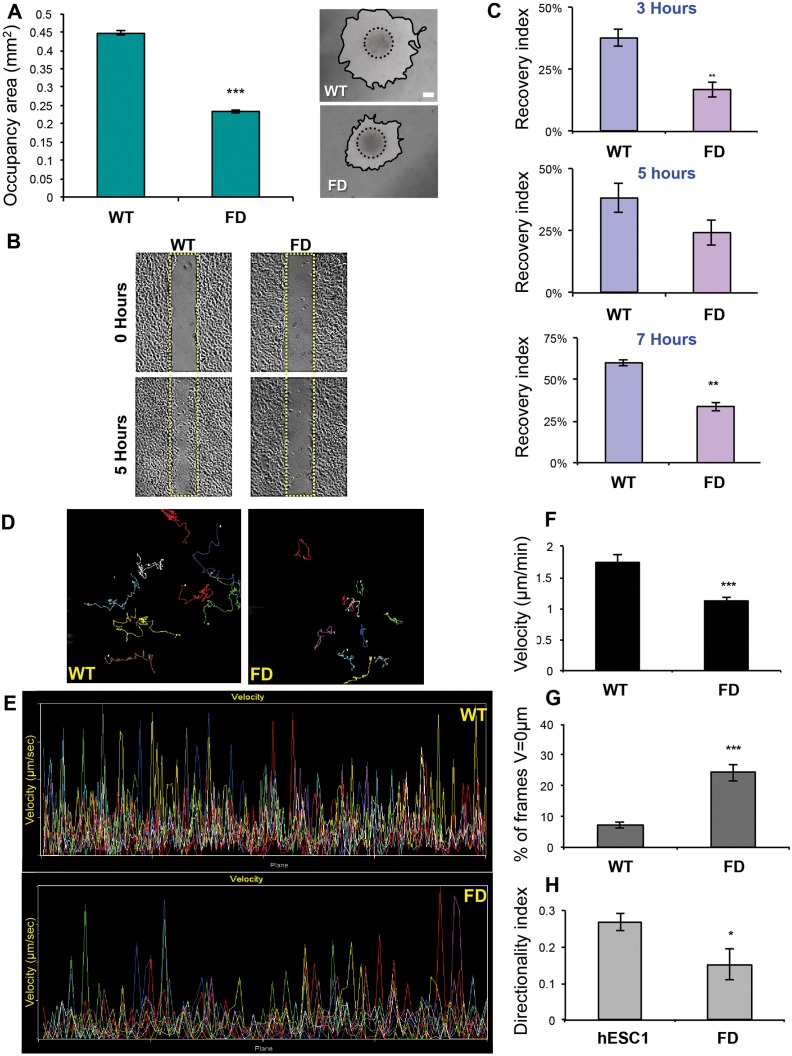
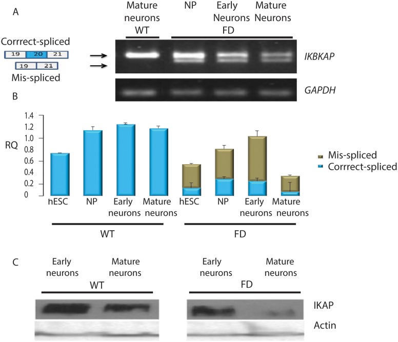
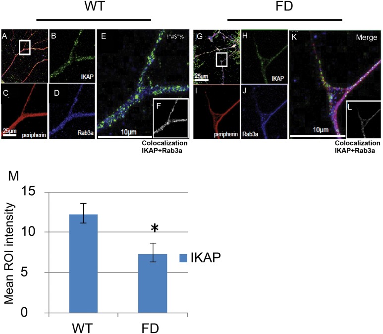
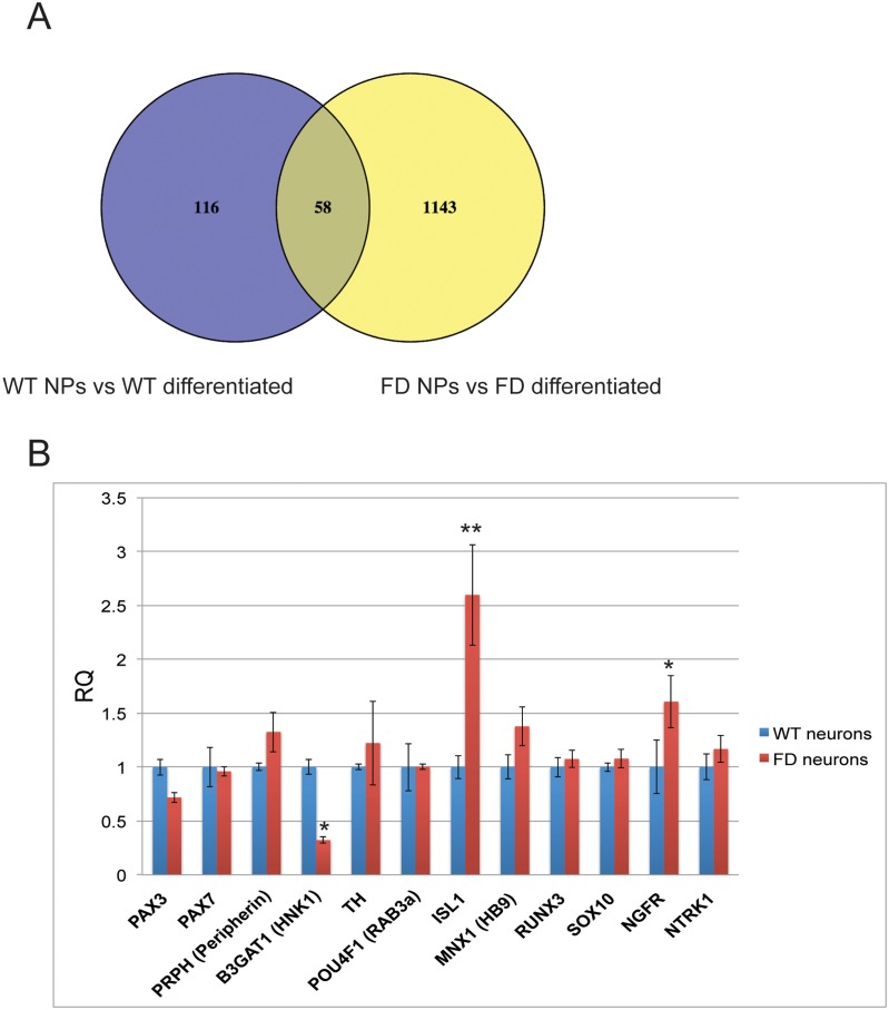
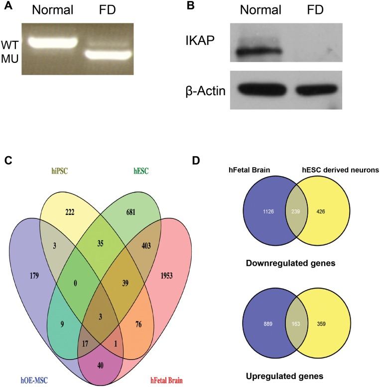
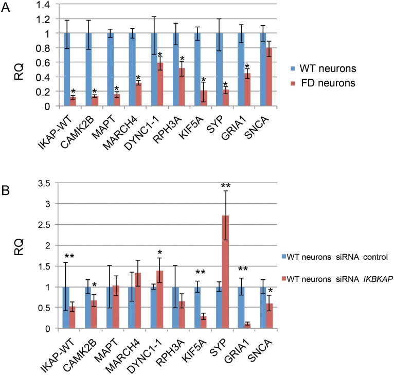
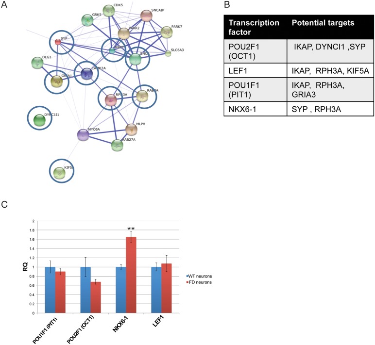
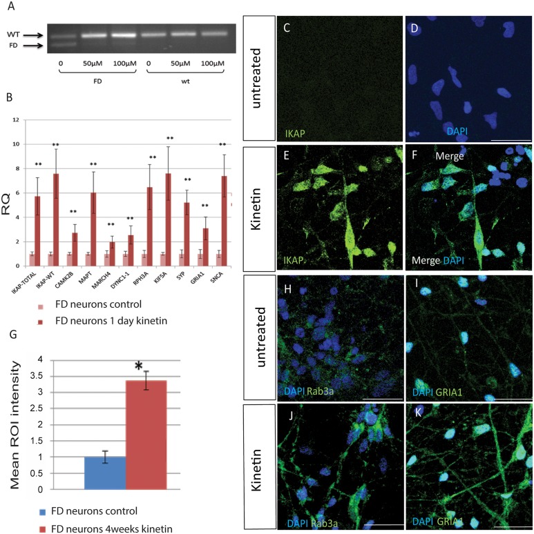
A splicing mutation in the IKBKAP gene causes Familial Dysautonomia (FD), affecting the IKAP protein expression levels and proper development and function of the peripheral nervous system (PNS). Here we found new molecular insights for the IKAP role and the impact of the FD mutation in the human PNS lineage by using a novel and unique human embryonic stem cell (hESC) line homozygous to the FD mutation originated by pre implantation genetic diagnosis (PGD) analysis. We found that IKBKAP downregulation during PNS differentiation affects normal migration in FD-hESC derived neural crest cells (NCC) while at later stages the PNS neurons show reduced intracellular colocalization between vesicular proteins and IKAP. Comparative wide transcriptome analysis of FD and WT hESC-derived neurons together with the analysis of human brains from FD and WT 12 weeks old embryos and experimental validation of the results confirmed that synaptic vesicular and neuronal transport genes are directly or indirectly affected by IKBKAP downregulation in FD neurons. Moreover we show that kinetin (a drug that corrects IKBKAP alternative splicing) promotes the recovery of IKAP expression and these IKAP functional associated genes identified in the study. Altogether, these results support the view that IKAP might be a vesicular like protein that might be involved in neuronal transport in hESC derived PNS neurons. This function seems to be mostly affected in FD-hESC derived PNS neurons probably reflecting some PNS neuronal dysfunction observed in FD.


Conflict of interest statement

Competing Interests: The authors have declared that no competing interests exist.

Figures

Fig 1
Fig 1. Analysis of FD-NCC migration.
Delamination and migration of cells were monitored 24 hours after NP clusters attachment on Fibronectin. Migratory cells represent a homogenous population of NCC (See S4 Fig). Measurements of the areas occupied by the migrating NCC 24 hours after cluster attachment revealed that the FD-NCC had limited ability of delamination and migration. Representing phase contrast images of migration from plated clusters of WT and FD hNP clusters are presented on the right panel (A). Wound healing assay also showed reduced migration of FD-NCC. Representing phase contrast images of WT and FD-NCC cultures at time 0 and 5h after pipette tip scratching are shown in (B; Yellow rectangles represent original scratched area at time 0). FD-NCC have significantly reduced migration ability as indicated by their lower recovery index 3, 5 and 7 hours after scratching of the cultures (C). Time-lapse microscopy was later used to investigate FD-NCC migration in single cells level. The migration path of each WT and FD-NCC cell was traced and represented by different colored line, allow the follow-up of the migration path of each single cell (D). Monitoring of the velocity of each single cell is presented (each in specific color) on individually time frame during the assay (E). The measured mean migration velocity of FD-NCC is significantly reduced compared to WT-NCC (F). The mean percentage of time frames in which the FD-NCC were not migrating is significantly higher compared to WT-NCC (G). Finally, the measured directionality index of FD-NCC was significantly reduced as well (H). * P<0.05;** P<0.01; *** P<0.001; Scale bars A-B 200μm.
Fig 2
Fig 2. IKAP expression in WT and FD early and mature neurons.
(A) RT-PCR analysis of the expression of IKBKAP showing WT (upper lane) and FD (mis-spliced, lower lane) mRNA isoforms at the stage of early neuronal precursors, early and mature neurons. (B) qRT-PCR analysis of the levels of IKBKAP WT (blue) and FD (green) mRNA isoforms. (C) Western blot analysis of IKAP protein levels in WT and FD early and mature neurons. Actin served as loading control.
Fig 3
Fig 3. Localization of IKAP in hESC-derived PNS neurons in respect to vesicular neuronal markers SV2 and Rab3a.
Confocal micrographs of hESC derived PNS neurons double-stained with Rabbit anti-hIKAP antibodies combined with either antibodies against synaptic vesicular markers, synaptic vesicle 2 (SV2) (A-G) or with Rab3a (H-J). (A-D) Low magnification images showing the IKAP and SV2 coexpression within neurites. (E-G) Magnification of a neurite exhibiting granular expression of IKAP and SV2. (F and G) High magnification of boxed area in E, showing partial colocalization of the two proteins (indicated by arrowheads in G) within distinct vesicle structures. (H) Low magnification image showing IKAP and Rab3a localization within the soma and along neurites. (I and J) High magnification of boxed area in H exhibiting partial protein colocalization pattern within distinct vesicles (indicated by arrowheads in J). Scale bars are indicated according to the magnification in each image.
Fig 4
Fig 4. Characterization of IKAP localization in PNS WT and FD hESC-derived cultured neurons.
Immunofluorescence confocal microscopy analysis was performed as shown in Fig 3. IKAP together with peripherin and Rab3a expression are shown within hESC derived neurons in WT (A-F) and FD (G-L) genetic backgrounds. Images B and H show the expression and localization of IKAP in WT and FD derived-neurons respectively. C and I, D and J show the expression of peripherin and Rab3a respectively. IKAP and Rab3a co-localization levels are shown in WT (F) and FD (L). Quantitative analysis of the mean intensity of IKAP and RAB3a colocalization levels in WT and FD axons is shown in M. 3D stacks of sequential confocal images were de-convolved using Huygens (scientific volume imaging-SVI) software and the analyzed for colocalization between the signals was performed using Imaris (Bitplan). Bar sizes are indicated in representative images. Mouse anti-hIKAP (BD Biosciences) was used in these experiments.
Fig 5
Fig 5. Comparative analysis of expression differences in WT and FD hESC derived PNS differentiation process.
(A) Venn diagram comparing the overall number of genes that showed > 2 fold expression differences between hNP and fully differentiated hESC-derived neurons in WT (in blue) and FD (in yellow) backgrounds. (B) The normalized relative quantification expression values are shown as mean ± s.d. of several developmental PNS markers and transcription factors in WT and FD hESC-derived neurons as obtained in the transcriptome cDNA chip analysis. * P<0.05 **P<0.01.
Fig 6
Fig 6. Comparative transcriptome analysis between human 12 weeks fetal WT and FD brains to WT and FD hESC derived neurons.
Prior to cDNA microarray chip analysis mRNA and protein extracts from the brain samples were used to validate the FD phenotype of the FD splicing mutation in the IKBKAP gene at the transcriptional and translational levels by: (A) RT-PCR analysis of the expression of IKBKAP in human 12 weeks fetal WT and FD brains showing WT (upper lane) and FD (mis-spliced, lower lane) mRNA isoforms in the FD brain only. (B) Western blot analysis of IKAP protein levels in WT and FD brains. Note the almost complete absence of IKAP in the FD brain. β-Actin served as loading control. Comparative transcriptome analysis between data obtained from both cDNA microarray chips of WT and FD fetal brains and hESC-derived PNS neurons was performed by cross-referencing genes which their expression levels differ significantly (>2 fold) between FD and WT. (C) Venn Diagram representation of a wide genome transcriptome analysis of common genes that are differentially expressed in FD-hESC-derived PNS neurons (in green) and in two other known FD stem cell neural-derived models (FD fibroblasts derived iPSC (in yellow) [23] and FD-hOE-MSC (in blue) [24] and in FD Fetal brain (in red). Cross-referencing genes with their expression levels difference of >2 fold change for hESC p<0.05 or >1.5fold change p<0.05 for iPSC and hOE-MSC between WT and FD were considered for analysis. The number at the crossection between the diagrams represents the number of genes that are shared by these multiple analyses. For the list of genes see S3 Table. (D) Gene sets were divided into downregulated and upregulated genes in both hESC-derived PNS neurons and Fetal brain biological systems. Venn diagram represents the results from this analysis showing in blue, the number of genes that differ in WT and FD in the fetal in vivo and in yellow, the number of genes that differ in WT and FD hESC derived neurons in vitro. The crossection between the diagrams represent the number of genes that are shared by both analyses. Upper diagrams represent the downregulated genes while the lower ones represent the upregulated genes.
Fig 7
Fig 7. qRT-PCR analysis for validation of IKBKAP predicted co-regulated functional candidate genes in FD neurons and in siRNA IKBKAP downregulated WT neurons.
Validation experiments for IKBKAP predicted co-regulated functional candidate genes selected using the “Expander” micro arrays analysis tool were performed by qRT-PCR analysis on cDNA produced from FD hESC derived PNS neurons and on siRNA IKBKAP downregulated WT hESC derived PNS neurons (A) qRT-PCR results of relative quantification levels of IKBKAP and “candidate genes” in WT vs FD hESC derived PNS neurons. (B) qRT-PCR results of relative quantification levels of IKBKAP and “candidate genes” in WT-hESC derived PNS neurons that were treated with control siRNA and siRNA IKBKAP represented as mean ± s.d. * P<0.05 **P<0.01, Non statistical significance (NS).
Fig 8
Fig 8. Prediction analysis of networks and transcription factors regulating IKBKAP and IKBKAP co-regulated functional candidate genes.
“String” analysis of protein networks showing the potential interaction between the majority of the IKBKAP co-regulated functional candidate genes. (B) Prediction of transcription factors (TF) related to IKBKAP and co-regulated functional candidate genes. (C) Shows relative quantification levels represented as mean ± s.d. of TF gene candidates taken from the total cDNA microarray analysis showing difference between WT and FD-hESC derived PNS neurons. * P<0.05 **P<0.01, Non statistical significance (NS).
Fig 9
Fig 9. Rescue effect of kinetin on IKBKAP expression and concomitantly on in FD-hESC derived PNS neurons.
(A) RT-PCR results of one day kinetin treatment on FD-hESC derived PNS neurons showing the drug correcting effect on FD alternative splicing. (B) qRT-PCR analysis of the relative quantification levels represented as mean ± s.d. of IKBKAP and co-regulated functional candidates in FD neurons following one day kinetin treatment. * P<0.05 **P<0.01. (C-K) Effect of 4 weeks kinetin treatment on the expression of IKAP in FD-hESC derived PNS neurons (C-D, untreated and E-F, 4 weeks kinetin treated by immunofluorescence confocal analysis. (G) Quantification of IKAP mean levels calculated from mean RGB after 4 weeks kinetin treatment on these cells (C-K). * P<0.05. (H-K) Effect of 4 weeks kinetin treatment (J and K) on the expression of Rab3a (H and J) and GRIA1 (I and K) in FD-hESC derived PNS neurons by immunofluorescence confocal analysis. Mouse anti-hIKAP antibody (Abnova Corporation) was used in these experiments.

Similar articles

Cited by

References

    1. Riley CM, Day RL, Greeley DM, Langford WS. Central autonomic dysfunction with defective lacrimation: report of five cases. Pediatrics. 1949;3:468–78. - PubMed
    1. Axelrod FB. Familial dysautonomia. Muscle Nerve. 2004;29(3):352–63. 10.1002/mus.10499 - DOI - PubMed
    1. Blumenfeld A, Slaugenhaupt SA, Axelrod FB, Lucente DE, Maayan C, Liebert CB, et al. Localization of the gene for familial dysautonomia on chromosome 9 and definition of DNA markers for genetic diagnosis. Nat Genet. 1993;4(2):160–4. - PubMed
    1. Blumenfeld A, Slaugenhaupt SA, Liebert CB, Temper V, Maayan C, Gill SP, et al. Precise genetic mapping and haplotype analysis of the familial dysautonomia gene on human chromosome 9q31. Am J Hum Genet. 1999;64(4):1110–8. - PMC - PubMed
    1. Cuajungco MP, Leyne M, Mull J, Gill SP, Lu W, Zagzag D, et al. Tissue-specific reduction in splicing efficiency of IKBKAP due to the major mutation associated with familial dysautonomia. Am J Hum Genet. 2003;72(3):749–58. - PMC - PubMed

Publication types

MeSH terms