. 2015 Oct 6:5:14813.
doi: 10.1038/srep14813.

Antimicrobial activity of iron oxide nanoparticle upon modulation of nanoparticle-bacteria interface

Affiliations

Antimicrobial activity of iron oxide nanoparticle upon modulation of nanoparticle-bacteria interface

Manoranjan Arakha et al. Sci Rep. .

Abstract

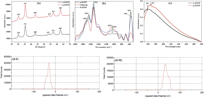
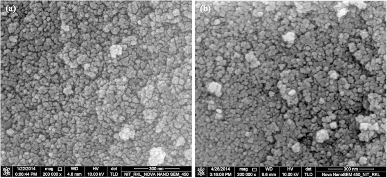
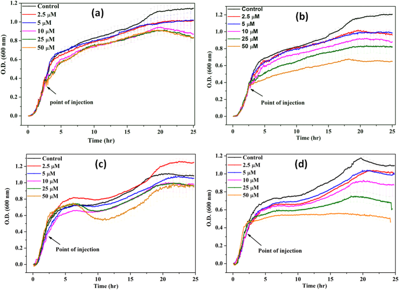
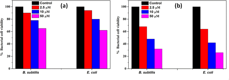
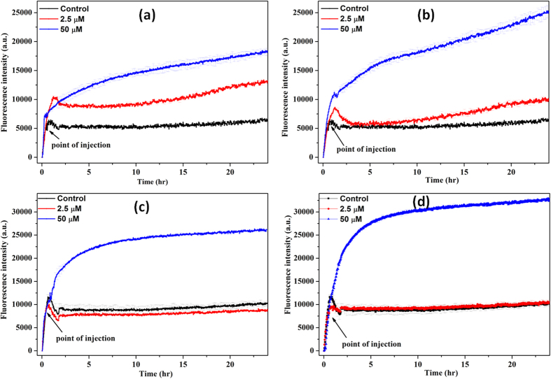
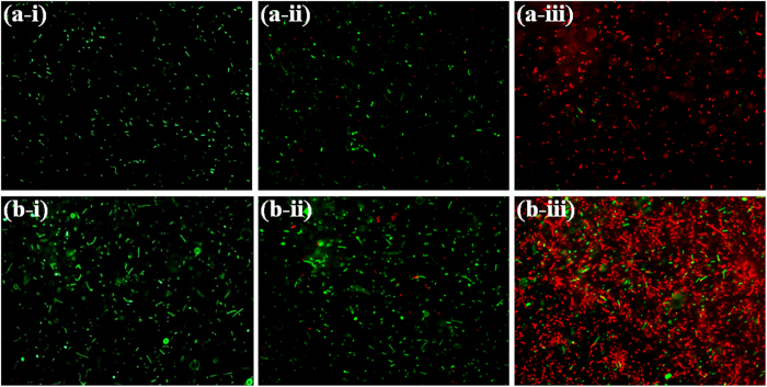
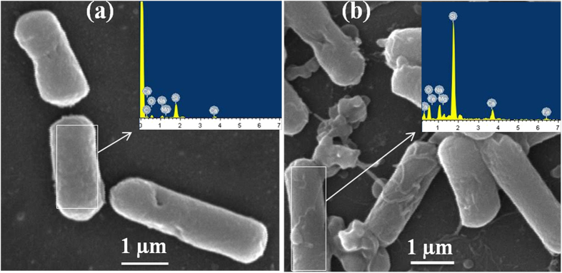
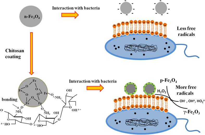
Investigating the interaction patterns at nano-bio interface is a key challenge for safe use of nanoparticles (NPs) to any biological system. The study intends to explore the role of interaction pattern at the iron oxide nanoparticle (IONP)-bacteria interface affecting antimicrobial propensity of IONP. To this end, IONP with magnetite like atomic arrangement and negative surface potential (n-IONP) was synthesized by co-precipitation method. Positively charged chitosan molecule coating was used to reverse the surface potential of n-IONP, i.e. positive surface potential IONP (p-IONP). The comparative data from fourier transform infrared spectroscope, XRD, and zeta potential analyzer indicated the successful coating of IONP surface with chitosan molecule. Additionally, the nanocrystals obtained were found to have spherical size with 10-20 nm diameter. The BacLight fluorescence assay, bacterial growth kinetic and colony forming unit studies indicated that n-IONP (<50 μM) has insignificant antimicrobial activity against Bacillus subtilis and Escherichia coli. However, coating with chitosan molecule resulted significant increase in antimicrobial propensity of IONP. Additionally, the assay to study reactive oxygen species (ROS) indicated relatively higher ROS production upon p-IONP treatment of the bacteria. The data, altogether, indicated that the chitosan coating of IONP result in interface that enhances ROS production, hence the antimicrobial activity.


Figures

Figure 1
Figure 1. Characterization of n-IONP and p-IONP.
(a) XRD spectra (b) ATR-FTIR absorption spectra, and (c) UV-Vis absorption spectra of n-IONP, and p-IONP, (d) Zeta potential analysis of n-IONP (Fig. d–I), and p-IONP (Fig. d–II).
Figure 2
Figure 2. FE-SEM image of n-IONP (Fig. 2a) and p-IONP (Fig. 2b).
Figure 3
Figure 3. Growth kinetics of B. subtilis (Figs. 3a and 3b) and E. coli (Figs. 3c and 3d) in absence and presence of different concentrations of n-IONP (Fig. 3a for B. subtilis and 3c for E. coli) & p-IONP (Fig. 3b for B. subtilis and 3d for E. coli).
Different concentrations of the NPs taken were 2.5, 5, 10, 25, and 50 μM, and injected at the log phase of growth kinetics (shown by arrow). Triplicate experiments were done for each reaction, and the error bar represents the standard error of mean.
Figure 4
Figure 4. Quantification of bacterial cell viability at different concentrations of n-IONP (Fig. 4a) and p-IONP (Fig. 4b).
Colony forming units (CFU) were quantified for both B. subtilis and E. coli cells, and represented as percentage of viable cells in comparison to colony obtained from untreated culture.
Figure 5
Figure 5. n-IONP and p-IONP induced ROS production.
Figure 5(a,c) represent change in fluorescence intensity with DCFH-DA oxidation in presence of n-IONP for B. subtilis and E. coli, respectively. Whereas figure 5(b,d) represent DCFH-DA oxidation kinetics in presence of p-IONP for B. subtilis and E. coli, respectively. Each curve represents the average of three independent measurements with corresponding standard error of mean.
Figure 6
Figure 6. Fluorescence microscopic images of B. subtilis and E. coli in absence and presence of n-IONP and p-IONP.
Intact B. subtilis (a-i), B. subtilis in presence of 50 μM of n-IONP (a-ii), and B. subtilis in presence of 50 μM of p-IONP (a-iii), intact E. coli (b-i), E. coli in presence of 50 μM of n-IONP (b-ii), and E. coli in presence of 50 μM of p-IONP (b-iii). The scale bars represent for 20 μm.
Figure 7
Figure 7. SEM micrographs showing membrane deformation/damage of B. subtilis upon p-IONP treatment.
(a) SEM image of control (without p-IONP treatment), and figure inset shows the EDX spectra of B. subtilis surface. (b) SEM image of B. subtilis cells upon p-IONP treatment, and figure inset shows the EDX spectra of B. subtilis surface after p-IONP treatment.
Figure 8
Figure 8. Proposed schematic model elucidating the detail mechanism of IONPs against bacterial cells.

References

    1. Inbaraj B. S., Tsai T.-Y. & Chen B.-H. Synthesis, characterization and antibacterial activity of superparamagnetic nanoparticles modified with glycol chitosan. Sci Technol Adv Mater 13, 015002 (2012). - PMC - PubMed
    1. Dwivedi S. et al. Reactive oxygen species mediated bacterial biofilm inhibition via zinc oxide nanoparticles and their statistical determination. PLoS One 9, 10.1371/journal.pone.0111289 (2014). - DOI - PMC - PubMed
    1. Kaittanis C., Nath S. & Perez J. M. Rapid nanoparticle-mediated monitoring of bacterial metabolic activity and assessment of antimicrobial susceptibility in blood with magnetic relaxation. PLoS One 3, 10.1371/journal.pone.0003253 (2008). - DOI - PMC - PubMed
    1. Bjarnsholt T. et al. Why chronic wounds will not heal: a novel hypothesis. Wound Repair Regen 16, 2–10 (2008). - PubMed
    1. Nel A. E. et al. Understanding biophysicochemical interactions at the nano–bio interface. Nat Mater 8, 543–557 (2009). - PubMed

Publication types

MeSH terms