. 2015 Oct 6:5:14778.
doi: 10.1038/srep14778.

PLD1 participates in BDNF-induced signalling in cortical neurons

Affiliations

PLD1 participates in BDNF-induced signalling in cortical neurons

Mohamed Raafet Ammar et al. Sci Rep. .

Abstract

The brain-derived neurotrophic factor BDNF plays a critical role in neuronal development and the induction of L-LTP at glutamatergic synapses in several brain regions. However, the cellular and molecular mechanisms underlying these BDNF effects have not been firmly established. Using in vitro cultures of cortical neurons from knockout mice for Pld1 and Rsk2, BDNF was observed to induce a rapid RSK2-dependent activation of PLD and to stimulate BDNF ERK1/2-CREB and mTor-S6K signalling pathways, but these effects were greatly reduced in Pld1(-/-) neurons. Furthermore, phospho-CREB did not accumulate in the nucleus, whereas overexpression of PLD1 amplified the BDNF-dependent nuclear recruitment of phospho-ERK1/2 and phospho-CREB. This BDNF retrograde signalling was prevented in cells silenced for the scaffolding protein PEA15, a protein which complexes with PLD1, ERK1/2, and RSK2 after BDNF treatment. Finally PLD1, ERK1/2, and RSK2 partially colocalized on endosomal structures, suggesting that these proteins are part of the molecular module responsible for BDNF signalling in cortical neurons.


Figures

Figure 1
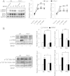
Figure 1. PLD1 and RSK2 expression and PLD activity in cultured cortical neurons.
(A) E17 cortical neurons from control C57BL6 mice were cultured and lyzed between 3 and 12 DIV. 35 μg of proteins in each condition was resolved by SDS-PAGE and probed with anti-PLD1, anti-RSK2 and anti-GAPDH antibodies. The expression levels of PLD1 and RSK2 were quantified for two independent experiments and normalized to GAPDH levels. (B) WT and Rsk2−/− cortical neurons at 3 DIV were incubated for 1 to 60 minutes with 100 ng/mL of BDNF and used to measure PLD activity. Data are normalized to the activity in WT neurons in the absence of treatment and were obtained from two independent measurements with sextuplicates.
Figure 2
Figure 2. PLD1 is involved in different signalling pathway triggered by BDNF in cultured cortical neurons.
(A) WT and Pld1−/− cortical neurons in culture for 3 DIV were treated for the indicated time with 100 ng/mL of BDNF and lyzed. 35 μg of proteins was resolved by SDS-PAGE and probed with anti-ERK1/2, anti-phospho-ERK1/2 and anti-GAPDH antibodies. The phosphorylation level of ERK1/2 was corrected to ERK1/2 expression levels and subsequently normalized to GAPDH expression levels. The relative ERK1 and ERK2 is normalized to that of WT untreated cells obtained from three independent experiments. (B) WT and Pld1−/− cortical in culture for 3 DIV were treated for 15 minutes with 100 ng/mL of BDNF and lyzed. 35 μg of proteins was resolved by SDS-PAGE and probed with anti-phospho-mTor (Ser-2481), anti-phospho-S6K (Thr-389), anti-phospho-CREB (Ser-133) and anti-GAPDH antibodies. The levels of phospho-proteins was normalized to GAPDH expression levels and normalized to that of WT untreated cells. For pS6K quantification, lower bands levels corresponding to P70S6K were analysed. Data were averaged from three independent experiments.
Figure 3
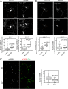
Figure 3. PLD1 regulates the nuclear level of p-ERK1/2 and p-CREB.
2 DIV WT cortical neurons were transfected with PLD1-GFP and after 24 hours treated for 15 minutes with 100 ng/mL of BDNF. After fixation cells were stained with anti-phospho-ERK1/2 (A) or anti-phospho-CREB (B) antibodies and the nucleus stained with DAPI (not shown). (C) WT and Pld1−/− cortical neurons in culture for 3 DIV were treated for 15 minutes with 100 ng/mL of BDNF. After fixation cells were stained with anti-phospho-CREB and anti-β-tubulin antibodies, and the nucleus was stained with DAPI (not shown). The level of nuclear phospho-ERK1/2 (A) or phospho-CREB (B,C) fluorescence intensity was normalized to the nuclear area revealed by DAPI staining from at least 30 cells per condition. Bar = 25 μm.
Figure 4
Figure 4. PLD1 colocalizes with p-ERK1/2 and RSK2 on vesicular structures.
2 DIV WT cortical neurons were transfected with PLD1-GFP (A) or PLD1-GFP and HA-RSK2 and 24 hours later, treated for 15 minutes with 100 ng/mL BDNF. After fixation, cells were stained with anti-phospho-ERK1/2 and/or with anti-HA antibodies. Selected regions of interest revealed partial colocalization of PLD1-GFP with phospho-ERK1/2 (A) and of PLD1-GFP with HA-RSK2, and with phospho-ERK1/2 (B). Similar observations were obtained from two independent cultures. Bars = 25 μm.
Figure 5
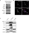
Figure 5. PLD1, p-ERK1/2 and PEA15 are found on late endosomal vesicles.
(A) 3 DIV neurons treated for 15 minutes with 100 ng/mL of BDNF were lyzed and subjected to subcellular fractionation by velocity centrifugation on OptiPrep gradient (5-10-15-20-25%). Fractions were collected and after Western blotting probed with anti-TrkB, anti-PLD1, anti-APPL1, anti-RSK2, anti-pERK1/2, anti-Rab5, and anti-PEA15 antibodies. Similar observations were obtained on two independent experiments. (B) 2 DIV WT cortical neurons were transfected with PLD1-GFP and 24 hours later treated for 15 minutes with 100 ng/mL BDNF. After fixation, cells were stained with anti-phospho-ERK1/2 and anti-Rab7 antibodies. Arrows indicate vesicular structures positives for PLD1, Rab7, and phospho-ERK1/2. Bar = 25 μm. (C) 3 DIV neuronal cultures untreated (−) or treated ( + ) for 15 minutes with 100 ng/mL BDNF were lyzed, and PEA15 was immunoprecipitated. Pre-immunoprecipitation, immunoprecipitated and post-immuniprecipitated samples were probed with anti-PEA15, anti-PLD1, anti-RSK2, anti-ERK1/2, and anti-GAPDH antibodies. GAPDH was used a negative control of immuonprecipitation. Note that the amount of PEA15 immunoprecipitated after BDNF treatment varied and that the amount of ERK1/2 coimmunoprecipitated varied accordingly. Similar observations were replicated twice.
Figure 6
Figure 6. Effect of PEA15 silencing on BDNF-induced increases in the nuclear level of p-ERK1/2 and p-CREB.
(A) 2 DIV WT cortical neurons were transfected with two different siRNA for PEA15, an unrelated siRNA, or mock-transfected. After 72 h, cells were lysed and 35 μg of proteins was resolved by SDS-PAGE and probed with anti-PEA15 and anti-GAPDH antibodies. Similar results were obtained in two independent experiments. (B,C) Cortical neurons expressing either the unrelated siRNA, or PEA15-siRNA#1, or PEA15-siRNA#2, were untreated (−) or treated ( + ) for 15 minutes with 100 ng/mL of BDNF. After fixation, cells were stained with anti-phospho-ERK1/2 or anti-phospho-CREB antibodies and the nucleus stained with DAPI (not shown). The level of nuclear phospho-ERK1/2 (B) or phospho-CREB (C) fluorescence intensity was normalized to the nuclear area revealed by DAPI staining.

References

    1. Gottmann K., Mittmann T. & Lessmann V. BDNF signaling in the formation, maturation and plasticity of glutamatergic and GABAergic synapses. Exp Brain Res 199, 203–234, 10.1007/s00221-009-1994-z (2009). - DOI - PubMed
    1. Park H. & Poo M. M. Neurotrophin regulation of neural circuit development and function. Nat Rev Neurosci 14, 7–23, 10.1038/nrn3379 (2013). - DOI - PubMed
    1. Zagrebelsky M. & Korte M. Form follows function: BDNF and its involvement in sculpting the function and structure of synapses. Neuropharmacology 76 Pt C, 628–638, 10.1016/j.neuropharm.2013.05.029 (2014). - DOI - PubMed
    1. McAllister A. K. Spatially restricted actions of BDNF. Neuron 36, 549–550 (2002). - PubMed
    1. Horch H. W. & Katz L. C. BDNF release from single cells elicits local dendritic growth in nearby neurons. Nat Neurosci 5, 1177–1184, 10.1038/nn927 (2002). - DOI - PubMed

Publication types

MeSH terms

Substances

LinkOut - more resources