. 2015 Oct 6:12:30.
doi: 10.1186/s12989-015-0105-5.

Effects of prenatal inhalation exposure to copper nanoparticles on murine dams and offspring

Affiliations

Effects of prenatal inhalation exposure to copper nanoparticles on murine dams and offspring

Andrea Adamcakova-Dodd et al. Part Fibre Toxicol. .

Abstract

Background: Increasing numbers of individuals may be exposed to nanomaterials during pregnancy. The overarching goal of this investigation was to determine if prenatal inhalation exposure to copper nanoparticles (Cu NPs) has an effect on dams and offspring, including an analysis of inflammatory markers (Th1/Th2 cytokine profiles).

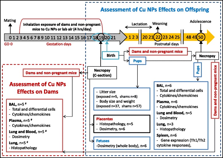
Methods: Physicochemical characterization of Cu NPs was performed. Pregnant and non-pregnant mice (C57Bl/6 J) were exposed to Cu NPs or laboratory air in the whole-body chamber for 4 hrs/day on gestation days (GD) 3-19 (3.5 mg/m(3)). Animals were euthanized on GD 19 (0 week) or 7 weeks later. Bronchoalveolar lavage (BAL) fluid was analyzed for total and differential cells. Cytokine/chemokine concentrations were determined in the BAL fluid and the plasma of dams/non-pregnant mice and pups. Cu content was determined in the lungs and the blood of dams/non-pregnant mice and pups, in the placentas as well as in the whole bodies of pups immediately after delivery. Lungs and placentas were evaluated for histopathological changes. Gene expression of the Th1/Th2 profiles were analyzed in spleens of pups.

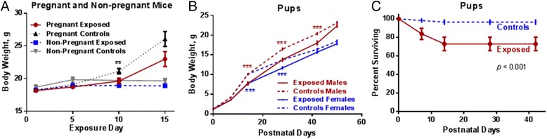
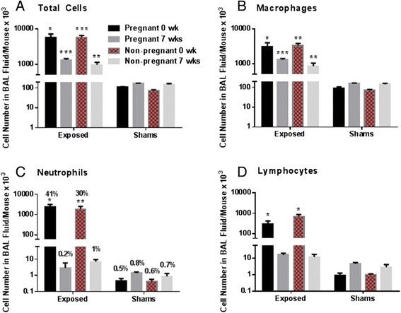
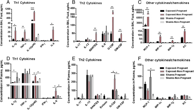
Results: The survival rate of 7 week old pups exposed to Cu NPs was significantly lower than control pups (73 vs. 97 %). The average litter size, male/female ratio, body weight and lenght at birth were not different between Cu NP-exposed and control mice. Both pregnant and non-pregnant mice exposed to Cu NPs had significant pulmonary inflammation with increased number of neutrophils in the BAL fluid compared to controls. Perivascular lymphoplasmacytic cuffing was found in the lungs of exposed mice and was more pronounced in the non-pregnant group. Similarly, levels of inflammatory cytokines/chemokines IL-12(p40), G-CSF, GM-CSF, KC, MCP-1, MIP-1α, MIP-1β, RANTES and TNF-α in BAL fluid were significantly higher in non-pregnant than pregnant exposed mice. Histopathology evaluation of placentas did not identify any pathological changes. No translocation of Cu into the placenta or the fetus was found by inductively coupled plasma-mass spectroscopy. Expression of several Th1/Th2 or other immune response genes in pups' spleens were found to be significantly up- or down-regulated.

Conclusions: Prenatal exposure to Cu NPs caused a profound pulmonary inflammation in dams and strong immunomodulatory effects in offspring. There was no clear polarization of genes expressed in pups' spleens towards Th1 or Th2 type of response.


Figures

Fig. 1
Fig. 1
The experimental study design and the number of animals per each evaluated endpoint. All mice (dams and non-pregnant mice) were exposed to Cu NPs or filtered laboratory air (4 hrs/day) from gestation day (GD) 3 to 19. A selected number of mice was euthanized on GD 19 and the pups were delivered by Caesarian section (0 wk post exposure). Another group of mice delivered the pups spontaneously on GD 18–20, these pups grew until adolescence when they were euthanized at 50 postnatal days (PND). # There were 4 mice in the pregnant group exposed to Cu NPs, all other experimental groups had 5 animals
Fig. 2
Fig. 2
Characterization of Cu NPs. Particle size distribution of primary Cu NPs (a) TEM image of primary Cu NPs (b), and the size distribution of generated Cu NP aerosol in the whole-body exposure chamber (c)
Fig. 3
Fig. 3
Body weights of all animals and survival rate of pups. The body weights of pregnant and non-pregnant mice during Cu NPs exposure (a). Postnatal body weights of pups (b) and postnatal survival rate of pups after prenatal inhalation exposure to Cu NPs (c). Significant value **p < 0.1 in a represents the comparison between pregnant exposed vs. pregnant controls. Significant value ***p < 0.001 in b represents the comparison between exposed males vs. control males and exposed females vs. control females pups
Fig. 4
Fig. 4
Cell numbers in BAL fluid of exposed and control pregnant and non-pregnant mice. A comparison of the number of total cells (a), macrophages (b), neutrophils (c) and lymphocytes (d) in the BAL fluid in pregnant and non-pregnant mice exposed to Cu NPs and their sham counterparts. c the percentage of neutrophils out of the total number of cells is also shown. Values are expressed as mean ± SE. Statistically significant differences between exposed groups and control counterparts (shams) are indicated as follows: *p < 0.05, **p < 0.01 and ***p < 0.001
Fig. 5
Fig. 5
Cell numbers in BAL fluid of exposed and control pups. The total cell number and number of macrophages, neutrophils and lymphocytes in the BAL fluid of mice at 50 PNDs that were exposed to Cu NPs during gestation days 3–19. Values are expressed as mean ± SE. Statistically significant difference between the exposed group and the control group is indicated as *p < 0.05
Fig. 6
Fig. 6
Cytokine/chemokine concentrations. The concentration of cytokines/chemokines in the BAL fluid (a-c) and the plasma (d-f) of pregnant and non-pregnant mice exposed to Cu NPs during gestation and their control counterparts divided into functional groups as follows: Th1 cytokines (a and d), Th2 cytokines (b and e) and other cytokines/chemokines (c and f). Values are expressed as mean ± SE. Statistically significant differences between the exposed groups and the control counterparts (shams) are indicated as follows: *p < 0.05, **p < 0.01 and ***p < 0.001
Fig. 7
Fig. 7
Lung histopathology. Lung from mice exposed to Cu NPs and euthanized immediately after C-section on GD 19 (0 PND) exhibited histopathologic changes including mild to moderate, multifocal inflammatory cell infiltration within the interstitium which is composed primarily of lymphocytes and neutrophils with fewer plasma cells and macrophages (thick arrows). Also present was multifocal accumulations of a basophilic, flocculent, amorphous material (thin arrows). The pathology findings were more pronounced in non-pregnant mice as opposed to the pregnant group. There were no pathologic findings in control mice. Bars = 200 μm (inset, bars = 20 μm)
Fig. 8
Fig. 8
Spleenic gene expression in pups. The mouse Th1 and Th2 responses were identified in RNAs isolated from the spleens of pups after prenatal exposure to Cu NPs by RT2 Profiler PCR Array (PAMM-034ZA, SABiosciences, Qiagen). Out of 84 genes, 14 genes were significantly (p < 0.05) up-regulated at least 2-fold and 11 genes were significantly (p < 0.05) down-regulated at least 2-fold from the controls

References

    1. Schwartz J. Air pollution and blood markers of cardiovascular risk. Environ Health Perspect. 2001;109(Suppl 3):405–9. doi: 10.1289/ehp.01109s3405. - DOI - PMC - PubMed
    1. Peters A, Liu E, Verrier RL, Schwartz J, Gold DR, Mittleman M, Baliff J, Oh JA, Allen G, Monahan K, Dockery DW. Air pollution and incidence of cardiac arrhythmia. Epidemiology. 2000;11(1):11–7. doi: 10.1097/00001648-200001000-00005. - DOI - PubMed
    1. Franck U, Odeh S, Wiedensohler A, Wehner B, Herbarth O. The effect of particle size on cardiovascular disorders - The smaller the worse. Sci Total Environ. 2011;409(20):4217–4221. doi: 10.1016/j.scitotenv.2011.05.049. - DOI - PubMed
    1. Pope CA, 3rd, Burnett RT, Thun MJ, Calle EE, Krewski D, Ito K, Thurston GD. Lung cancer, cardiopulmonary mortality, and long-term exposure to fine particulate air pollution. JAMA. 2002;287(9):1132–41. doi: 10.1001/jama.287.9.1132. - DOI - PMC - PubMed
    1. Sram RJ, Binkova B, Dejmek J, Bobak M. Ambient air pollution and pregnancy outcomes: a review of the literature. Environ Health Perspect. 2005;113(4):375–82. doi: 10.1289/ehp.6362. - DOI - PMC - PubMed

Publication types