. 2015 Oct 5:14:389.
doi: 10.1186/s12936-015-0922-x.

A potential role for interleukin-33 and γ-epithelium sodium channel in the pathogenesis of human malaria associated lung injury

Affiliations

A potential role for interleukin-33 and γ-epithelium sodium channel in the pathogenesis of human malaria associated lung injury

Sumate Ampawong et al. Malar J. .

Abstract

Background: The pathogenesis of pulmonary oedema (PE) in patients with severe malaria is still unclear. It has been hypothesized that lung injury depends, in addition to microvascular obstruction, on an increased pulmonary capillary pressure and altered alveolar-capillary membrane permeability, causing pulmonary fluid accumulation.

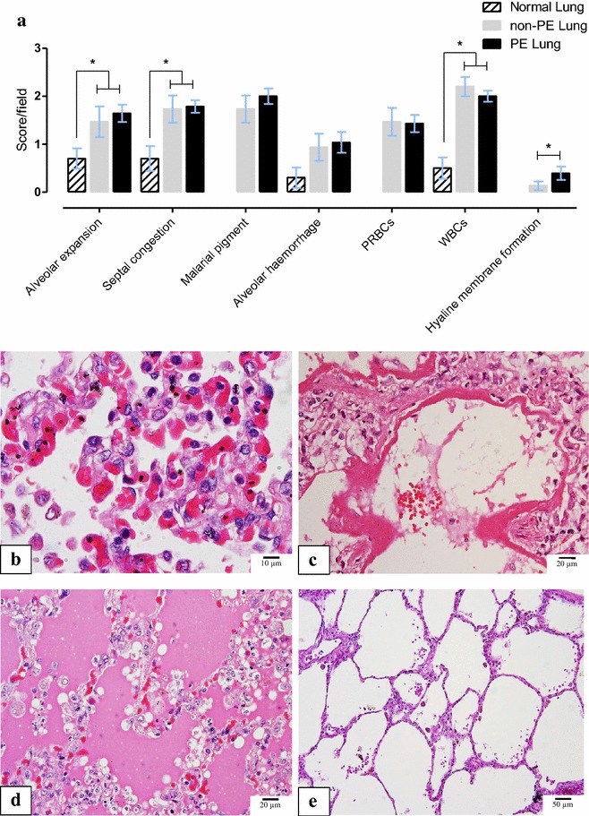
Methods: This study compared the histopathological features of lung injury in Southeast Asian patients (n = 43) who died from severe Plasmodium falciparum malaria, and correlated these with clinical history in groups with or without PE. To investigate the expression of mediators that may influence fluid accumulation in PE, immunohistochemistry and image analysis were performed on controls and sub-sets of patient with or without PE.

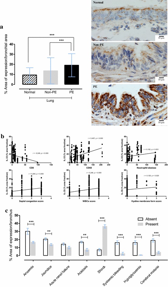
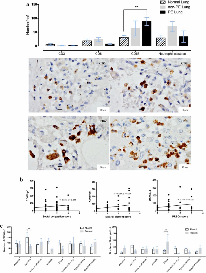
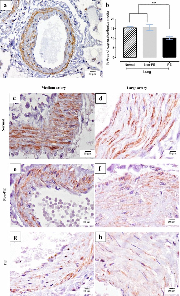
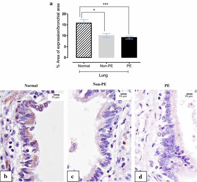
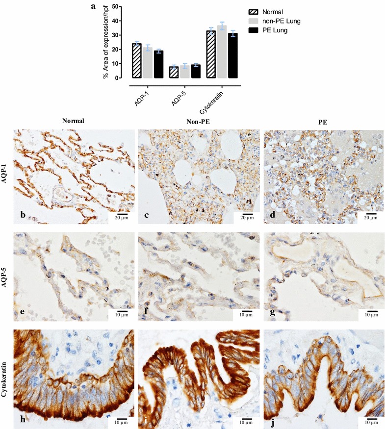
Results: The expression of leukocyte sub-set antigens, bronchial interleukin (IL)-33, γ-epithelium sodium channel (ENaC), aquaporin (AQP)-1 and -5, and control cytokeratin staining was quantified in the lung tissue of severe malaria patients. Bronchial IL-33 expression was significantly increased in severe malaria patients with PE. Malaria patients with shock showed significantly increased bronchial IL-33 compare to other clinical manifestations. Bronchial IL-33 levels were positively correlated with CD68+ monocyte and elastase + neutrophil, septal congestion and hyaline membrane formation. Moreover, the expression of both vascular smooth muscle cell (VSMC) and bronchial γ-ENaC significantly decreased in severe malaria patients with PE. Both VSMC and bronchial γ-ENaC were negatively correlated with the degree of parasitized erythrocyte sequestration, alveolar thickness, alveolar expansion score, septal congestion score, and malarial pigment score. In contrast AQP-1 and -5 and pan cytokeratin levels were similar between groups.

Conclusions: The results suggest that IL-33 may play a role in lung injury during severe malaria and lead to PE. Both VSMC and bronchial γ-ENaC downregulation may explain pulmonary fluid disturbances and participate in PE pathogenesis in severe malaria patients.


Figures

Fig. 1
Fig. 1
Histopathological changes in the lung in severe malaria. a Bar chart comparing score of the histopathological features in normal, non-PE and PE malaria lung samples; b, c photomicrographs of histopathological changes in a fatal falciparum malaria case [case number A41001]; b alveolar capillary expansion with accumulation of sequestration PRBCs and leukocytes accumulation. Phagocytosed haemozoin pigment in macrophages and neutrophils within alveolar septa; c pulmonary oedema and hyaline membrance formation indicated severe lung injury; d severe pulmonary oedema presented with free oedematous fluid in alveolar space [case number A47001-8]; and, e normal lung with clear alveolar space and thin alveolar septum [case number RC002-22], (H&E staining, scale bars as shown), For this and subsequent figures statistical significance of comparisons are *p < 0.05, **p < 0.01, ***p < 0.0001
Fig. 2
Fig. 2
Immunohistochemical expression of bronchial IL-33 expression is increased in malaria cases with pulmonary oedem. a Bar chart comparing of bronchial IL-33 expression in normal, non-PE and PE lung samples and photomicrographs of immunohistochemical staining of bronchial IL-33 localization; b significance correlation plot of bronchial IL-33 expression to leukocyte count and histopathological score; c bar chart comparing of bronchial IL-33 expression to clinical manifestations
Fig. 3
Fig. 3
Alveolar leukocyte sub-set counts compared between clinical groups and histopathological features. a Bar chart comparing of leukocyte sub-sets count in normal, non-PE and PE lung samples and photomicrographs of immunohistochemical staining of leukocyte sub-sets localization; b significance correlation plot of leukocyte sub-sets count to histopathological score; c significance bar chart comparing of leukocyte sub-sets count to clinical manifestations
Fig. 4
Fig. 4
Expression of vascular smooth muscle cell (VSMC) γ-ENaC. a Photomicrograph of small arteries tunica media’s VSMC show positive cytoplasmic immunostaining pattern; b Bar chart comparing of VSMC γ-ENaC expression in normal, non-PE and PE lung samples; photomicrographs of immunohistochemical staining of VSMC γ-ENaC in both medium-size and large arteries in normal (c, d), non-PE (e, f) and PE (g, h) lung samples
Fig. 5
Fig. 5
Immunohistochemical staining for bronchial γ-ENaC. a Bar chart comparing of bronchial γ-ENaC expression in normal, non-PE and PE lung samples; photomicrographs of immunohistochemical staining of bronchial γ-ENaC localization in normal (b), non-PE (c) and PE (d) lung samples
Fig. 6
Fig. 6
Immunohistochemical staining for AQP-1 and -5 and bronchial cytokeratin. a Bar chart comparing the degree of alveolar expression of AQP-1 and -5 and bronchial expression of cytokeratin in normal, non-PE, and PE-lung samples; photomicrographs of immunohistochemical staining of AQP-1 and -5 (localized on alveolar vessel and alveolar epithelium, respectively) and bronchial cytokeratin among normal (b, e, h), non-PE (c, f, i) and PE (d, g, j) lung samples

References

    1. White NJ, Ho M. The pathophysiology of malaria. Adv Parasitol. 1992;31:83–173. doi: 10.1016/S0065-308X(08)60021-4. - DOI - PubMed
    1. Taylor WR, Hanson J, Turner GD, White NJ, Dondorp AM. Respiratory manifestations of malaria. Chest. 2012;142:492–505. doi: 10.1378/chest.11-2655. - DOI - PubMed
    1. Taylor WR, White NJ. Malaria and the lung. Clin Chest Med. 2002;23:457–468. doi: 10.1016/S0272-5231(02)00004-7. - DOI - PubMed
    1. World Health Organiztion, Communicable Diseases Cluster Severe falciparum malaria. Trans R Soc Trop Med Hyg. 2000;94(Suppl 1):S1–S90. doi: 10.1016/S0035-9203(00)90300-6. - DOI - PubMed
    1. Aursudkij B, Wilairatana P, Vannaphan S, Walsh DS, Gordeux VR, Looareesuwan S. Pulmonary edema in cerebral malaria patients in thailand. Southeast Asian J Trop Med Public Health. 1998;29:541–545. - PubMed

Publication types

LinkOut - more resources