. 2015 Oct 4;5(12):2619-28.
doi: 10.1534/g3.115.019463.

HES-Mediated Repression of Pten in Caenorhabditis elegans

Affiliations

HES-Mediated Repression of Pten in Caenorhabditis elegans

Han Ting Chou et al. G3 (Bethesda). .

Abstract

The hairy/enhancer-of-split (HES) group of transcription factors controls embryonic development, often by acting downstream of the Notch signaling pathway; however, little is known about postembryonic roles of these proteins. In Caenorhabditis elegans, the six proteins that make up the REF-1 family are considered to be HES orthologs that act in both Notch-dependent and Notch-independent pathways to regulate embryonic events. To further our understanding of how the REF-1 family works to coordinate postembryonic cellular events, we performed a functional characterization of the REF-1 family member, HLH-25. We show that, after embryogenesis, hlh-25 expression persists throughout every developmental stage, including dauer, into adulthood. Like animals that carry loss-of-function alleles in genes required for normal cell-cycle progression, the phenotypes of hlh-25 animals include reduced brood size, unfertilized oocytes, and abnormal gonad morphology. Using gene expression microarray, we show that the HLH-25 transcriptional network correlates with the phenotypes of hlh-25 animals and that the C. elegans Pten ortholog, daf-18, is one major hub in the network. Finally, we show that HLH-25 regulates C. elegans lifespan and dauer recovery, which correlates with a role in the transcriptional repression of daf-18 activity. Collectively, these data provide the first genetic evidence that HLH-25 may be a functional ortholog of mammalian HES1, which represses PTEN activity in mice and human cells.

Keywords: dauer recovery; gene expression microarray; gonad morphology; hairy/enhancer-of-split; unfertilized oocytes.


Figures

Figure 1
Figure 1
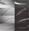
Postembryonic expression of Phlh-25::GFP. (A−D) Nomarski images (left) accompanied by green fluorescent protein fluorescent images (right). (A) Expression of cmjEx31 in multiple head neurons (arrowheads). This pattern was detected in animals at all stages, with a few additional neurons at dauer stage. (B) L3 expression in distal tip cell (arrow, red dashed circle), unidentified head neuron (arrowhead) and head muscles (asterisk). (C) L3 stage expression in PDE (arrow). Arrowheads indicate PDE processes. (D) Dauer stage expression in ventral nerve cord neurons. In all images anterior is to the right, dorsal is up. Scale bar represents 50 µm.
Figure 2
Figure 2
Brood size and unfertilized oocytes hlh-25(ok1710) animals. (A) hlh-25(ok1710) animals have fewer live progeny and lay more unfertilized eggs than wild-type animals. Expression of the extrachromosomal array cmjEx32, which carries hlh-25 genomic sequences, rescues this phenotype in hlh-25(ok1710) animals. Graph shows mean of total numbers of live progeny (dark bars) and unfertilized eggs (light bars) produced during the entire lifespans of wild-type (n = 7), hlh-25(ok1710) (n = 12), and hlh-25(ok1710; cmjEx32) animals (n = 9). *P-value < 0.05, **P-value < 0.005. (B) Average numbers of live progeny (solid lines) and unfertilized eggs (dashed lines) produced per day of egg laying by the same hlh-25(ok1710) animals (triangles) and wild-type animals (circles) as those shown in (A).
Figure 3
Figure 3
Gonad and oocyte morphologies of hlh-25(ok1710) animals. Nomarski (A, C) and merged fluorescent (B, D) images of representative gonad architecture and oocyte morphology in wild-type animals (A, B) as analyzed in a strain coexpressing a fluorescent chromosome marker (mCherry-Histone H2b) and a marker targeting green fluorescent protein to the plasma membrane (PI4,5P2) (Green et al. 2011), (B). Representative gonad architecture and oocyte morphology in hlh-25(ok1710) animals (C, D). Phenotypes include increased oocyte number, distal gonad arm constriction (arrow), increased apoptosis (^), and arrested embryos in uterus (#). Insert in (C) shows magnified view of distal gonad arm constriction. Inserts in (B) and (D) show embryonic nuclei. In all images, ventral is up, anterior is to the right, scale bar = 50 µm.
Figure 4
Figure 4
Functional annotation of HLH-25 targets. (A) Distribution of HLH-25 targets into top eight GO clusters. (B) Predicted and known interactions between genes in the ‘sexual reproduction’ cluster. The hub genes cgh-1, cyb-2.1, cyb-2.2, cyb-3, and puf-3 are outlined in red. The hub gene daf-18 is not a part of the ‘sexual reproduction’ cluster. (C) Representative genes from the 62 HLH-25 targets that form predicted or known interactions with daf-18. All genes, except daf-18, clustered under “embryonic development ending in birth (EDB)” Most genes also clustered under at least one other top GO category, including “nucleoside/nucleotide transport” (green spheres).
Figure 5
Figure 5
HLH-25 regulates lifespan and dauer recovery. (A) hlh-25(ok1710) animals (n = 282) have longer than lifespans than wild-type animals (n = 264, P-value < 0.0001). (B) Representative experiment showing the percentage of animals with pharyngeal pumping during dauer recovery at 20°. P-value < 0.0001 and P-value = 0.938 when hlh-25(ok1710) animals and daf-18(ok480) animals, respectively, were compared with wild-type. (C) Representative experiment showing the percentage of animals with pharyngeal pumping during dauer recovery at 20°. P-value < 0.0001 and P-value = 0.2771 when hlh-25(ok1710) and hlh-25(ok1710;cmjEx32) animals, respectively, were compared with wild-type.

References

    1. Akazawa C., Ishibashi M., Shimizu C., Nakanishi S., Kageyama R., 1995. A mammalian helix-loop-helix factor structurally related to the product of Drosophila proneural gene atonal is a positive transcriptional regulator expressed in the developing nervous system. J. Biol. Chem. 270: 8730–8738. - PubMed
    1. Alper S., Kenyon C., 2001. REF-1, a protein with two bHLH domains, alters the pattern of cell fusion in C. elegans by regulating Hox protein activity. Development 128: 1793–1804. - PubMed
    1. Audhya A., Hyndman F., McLeod I. X., Maddox A. S., Yates J. R., 3rd, et al. , 2005. A complex containing the Sm protein CAR-1 and the RNA helicase CGH-1 is required for embryonic cytokinesis in Caenorhabditis elegans. J. Cell Biol. 171: 267–279. - PMC - PubMed
    1. Axelson H., 2004. The Notch signaling cascade in neuroblastoma: role of the basic helix-loop-helix proteins HASH-1 and HES-1. Cancer Lett. 204: 171–178. - PubMed
    1. Ben-Arie N., Hassan B. A., Bermingham N. A., Malicki D. M., Armstrong D., et al. , 2000. Functional conservation of atonal and Math1 in the CNS and PNS. Development 127: 1039–1048. - PubMed

Publication types

MeSH terms

Substances