

Antenatal Three-Dimensional Printing of Aberrant Facial Anatomy
- PMID: 26438708
- PMCID: PMC4621796
- DOI: 10.1542/peds.2015-1062
Antenatal Three-Dimensional Printing of Aberrant Facial Anatomy
Abstract
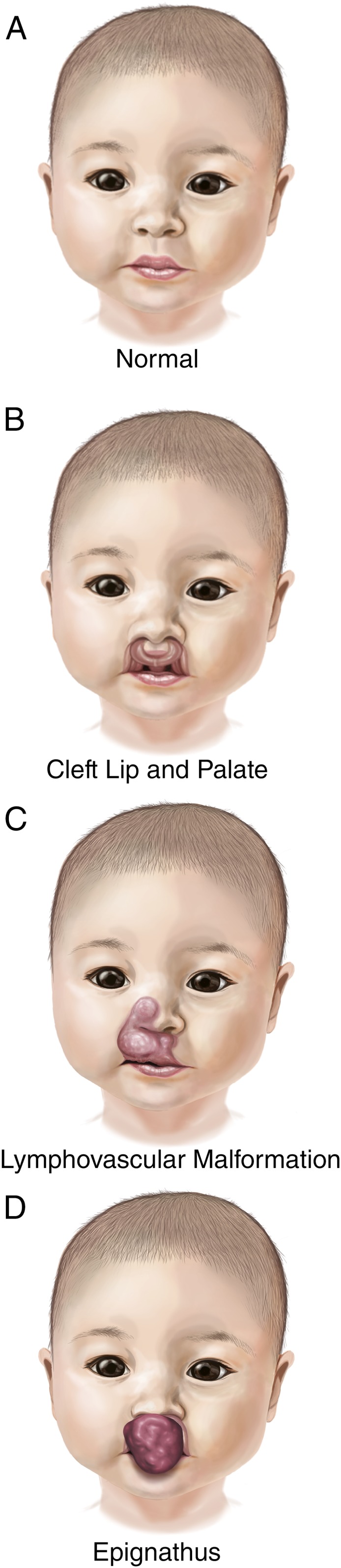
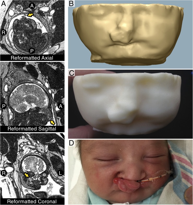
Congenital airway obstruction poses a life-threatening challenge to the newborn. We present the first case of three-dimensional (3D) modeling and 3D printing of complex fetal maxillofacial anatomy after prenatal ultrasound indicated potential upper airway obstruction from a midline mass of the maxilla. Using fetal MRI and patient-specific computer-aided modeling, the craniofacial anatomy of the fetus was manufactured using a 3D printer. This model demonstrated the mass to be isolated to the upper lip and maxilla, suggesting the oral airway to be patent. The decision was made to deliver the infant without a planned ex utero intrapartum treatment procedure. The neonate was born with a protuberant cleft lip and palate deformity, without airway obstruction, as predicted by the patient-specific model. The delivery was uneventful, and the child was discharged without need for airway intervention. This case demonstrates that 3D modeling may improve prenatal evaluation of complex patient-specific fetal anatomy and facilitate the multidisciplinary approach to perinatal management of complex airway anomalies.
Copyright © 2015 by the American Academy of Pediatrics.
Figures
References
-
- Hedrick MH, Ferro MM, Filly RA, Flake AW, Harrison MR, Adzick NS. Congenital high airway obstruction syndrome (CHAOS): a potential for perinatal intervention. J Pediatr Surg. 1994;29(2):271–274 - PubMed
-
- DeCou JM, Jones DC, Jacobs HD, Touloukian RJ. Successful ex utero intrapartum treatment (EXIT) procedure for congenital high airway obstruction syndrome (CHAOS) owing to laryngeal atresia. J Pediatr Surg. 1998;33(10):1563–1565 - PubMed
-
- Mychaliska GB, Bealer JF, Graf JL, Rosen MA, Adzick NS, Harrison MR. Operating on placental support: the ex utero intrapartum treatment procedure. J Pediatr Surg. 1997;32(2):227–230, discussion 230–231 - PubMed
-
- Coakley FV, Hricak H, Filly RA, Barkovich AJ, Harrison MR. Complex fetal disorders: effect of MR imaging on management—preliminary clinical experience. Radiology. 1999;213(3):691–696 - PubMed
-
- Mong A, Johnson AM, Kramer SS, et al. . Congenital high airway obstruction syndrome: MR/US findings, effect on management, and outcome. Pediatr Radiol. 2008;38(11):1171–1179 - PubMed
Publication types
MeSH terms
Grants and funding
LinkOut - more resources
Full Text Sources
Other Literature Sources
Medical
