. 2015 Dec;83(12):4861-70.
doi: 10.1128/IAI.00995-15. Epub 2015 Oct 5.

Brucella canis is an intracellular pathogen that induces a lower proinflammatory response than smooth zoonotic counterparts

Affiliations

Brucella canis is an intracellular pathogen that induces a lower proinflammatory response than smooth zoonotic counterparts

Carlos Chacón-Díaz et al. Infect Immun. 2015 Dec.

Abstract

Canine brucellosis caused by Brucella canis is a disease of dogs and a zoonotic risk. B. canis harbors most of the virulence determinants defined for the genus, but its pathogenic strategy remains unclear since it has not been demonstrated that this natural rough bacterium is an intracellular pathogen. Studies of B. canis outbreaks in kennel facilities indicated that infected dogs displaying clinical signs did not present hematological alterations. A virulent B. canis strain isolated from those outbreaks readily replicated in different organs of mice for a protracted period. However, the levels of tumor necrosis factor alpha, interleukin-6 (IL-6), and IL-12 in serum were close to background levels. Furthermore, B. canis induced lower levels of gamma interferon, less inflammation of the spleen, and a reduced number of granulomas in the liver in mice than did B. abortus. When the interaction of B. canis with cells was studied ex vivo, two patterns were observed, a predominant scattered cell-associated pattern of nonviable bacteria and an infrequent intracellular replicative pattern of viable bacteria in a perinuclear location. The second pattern, responsible for the increase in intracellular multiplication, was dependent on the type IV secretion system VirB and was seen only if the inoculum used for cell infections was in early exponential phase. Intracellular replicative B. canis followed an intracellular trafficking route undistinguishable from that of B. abortus. Although B. canis induces a lower proinflammatory response and has a stealthier replication cycle, it still displays the pathogenic properties of the genus and the ability to persist in infected organs based on the ability to multiply intracellularly.


Figures

FIG 1
FIG 1
Hematological profiles of infected dogs. The blood cell counts of 17 B. canis-infected dogs in a canine brucellosis outbreak in Costa Rica are shown. The gray area demarcates the normal value range of each cell type. The dogs are represented by circles numbered 1 to 17, with number 1 being the farthest to the left in each panel.
FIG 2
FIG 2
B. canis persists and replicates within cells of the reticuloendothelial system. Groups of 30 mice were inoculated i.p. with 107 CFU of B. canis-GFP or B. canis ΔvirB10-GFP. Groups of five mice were killed at the times indicated to determine the CFU counts in the spleen (A), liver (B), lymph nodes (C), and bone marrow (D). Cells from a spleen infected with B. canis-GFP for 21 days were visualized by epifluorescence (inset in panel A). Note the large amounts of B. canis-GFP within phagocytic cells. ΔvirB10-GFP was not observed within resident cells of the spleen. Error bars represent SDs. Data are representative of at least three independent experiments. Statistical significance was calculated by Student t test. *, P < 0.01.
FIG 3
FIG 3
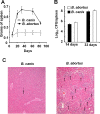
B. canis induces a lower proinflammatory response than B. abortus. Groups of 30 mice were inoculated i.p. with 107 CFU of B. canis-GFP or 106 CFU of B. abortus 2308. Groups of five mice were killed at various times to determine the spleen weight during 12 weeks of infection (A) and bacterial loads at 14 and 33 days p.i. (B). (C) Histological examination of the liver at 2 weeks p.i. with B. canis or B. abortus. Note that although the B. canis and B. abortus loads are similar (not statistically significantly different) at 2 weeks p.i., the granulomas (indicated by arrows) are more prominent in the B. abortus-infected liver than in the B. canis-infected liver. Error bars represent SDs. Data are representative of at least three independent experiments. In panel A, all of the values after 20 days are statistically significantly different (P < 0.001).
FIG 4
FIG 4
B. canis induces a lower cytokine response than B. abortus. Groups of 10 mice were inoculated i.p. with 107 CFU of B. canis-GFP or 106 CFU of B. abortus 2308. Groups of five mice were killed and bled after 2 and 3 weeks, respectively, to determine serum cytokine levels. Note the small amount of IFN-γ induced after 2 weeks by B. canis infection in comparison to that induced by B. abortus infection. The dashed lines represent the average background value (SD, <10%). Error bars represent SDs. Data are representative of at least three independent experiments. Statistical significance was calculated by Student t test. **, P < 0.001.
FIG 5
FIG 5
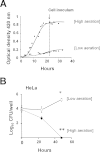
Modulation of the aeration conditions in the bacterial inoculum allows intracellular replication of B. canis. (A) B. canis cells (5 × 10⁹ CFU) were inoculated and grown for 30 h under high-aeration conditions (20 ml of TSB in 125-ml glass Erlenmeyer flasks, 37°C, 200 rpm) and low-aeration conditions (10 ml of TSB in 50-ml plastic tubes, 37°C, 120 rpm). Aliquots were taken at different times, and the optical densities at 420 nm were measured to determine the growth curves. (B) B. canis grown under the conditions indicated in panel A (dashed line) for 22 h was used to prepare the bacterial inoculum. HeLa cells were infected at an MOI of 500 in a gentamicin protection assay. After the incubation times indicated, CFU counts were determined. Error bars represent SDs. Data are representative of at least three independent experiments. Statistical significance was calculated by one-way analysis of variance. P values of <0.05 (*) and <0.01 (**) in relation to the corresponding T0 value of each bacterial condition are indicated.
FIG 6
FIG 6
The growth phase of the bacterial inoculum determines the ability of B. canis to replicate intracellularly. (A) B. canis cells (5 × 10⁹ CFU) were inoculated into 20 ml of TSB in a 125-ml Erlenmeyer flask and incubated at 37°C and 200 rpm for 30 h. Aliquots were taken at 5, 8, and 12 h (a, b, c), representing exponential-phase conditions, and at 24 and 30 h (d and e), representing stationary-phase conditions. O. D., optical density. Bacteria collected under each condition (a to e) were used to inoculate Raw 264.7 macrophages (MOI, 100 CFU) (B) and HeLa cells (MOI, 500 CFU) (C) in a gentamicin protection assay. At the times indicated, the number of CFU per well was determined. Error bars represent SDs. Data are representative of at least three independent experiments. In panels B and C, statistically significant differences between the 24- and 48-h points for each condition were calculated by Student t test. *, P < 0.01.
FIG 7
FIG 7
The B. canis virB10 mutant is unable to replicate in HeLa cells and Raw 264.7 macrophages. Early-exponential-phase (5 h) B. canis or B. canis ΔvirB10 was used to infect HeLa cells (MOI, 500 CFU) or Raw 264.7 macrophages (MOI, 100 CFU) in a gentamicin protection assay. At the times indicted, the number of CFU per well was determined. Error bars represent SDs. Data are representative of at least three independent experiments. Statistically significant differences between the counts achieved by both strains at 48 h were calculated by Student t test. *, P < 0.01.
FIG 8
FIG 8
B. canis displays two different patterns of interaction with epithelial cells. (A) HeLa cells were infected at an MOI of 500 CFU with an early-exponential-phase inoculum of B. canis-GFP grown under low-aeration conditions as indicated in Fig. 5A. After 48 h of incubation, cells were fixed and their nuclei were stained with 4′,6-diamidino-2-phenylindole (DAPI; blue) and visualized by fluorescence microscopy. (A) Two interacting patterns are shown: HeLa cells displaying scattered cell-associated bacteria (a to c) and intracellular replicating bacteria (d and e). (B) At 48 h p.i., living nonpermeabilized HeLa cells were incubated with an antibody to B. canis for 30 min at 4°C, followed by an anti-mouse antibody conjugated with Alexa Fluor 594 (Life Technologies). Cells were then fixed, permeabilized, and processed for an immunofluorescence assay. Intracellularly located bacteria are exclusively green (GFP signal), whereas extracellular bacteria are red (anti-B. canis signal). Images were contrasted and saturated with the Hue tool to obtain suitable color separation. Scale bars, 5 μm.
FIG 9
FIG 9
Intracellular B. canis bacteria are viable replicating bacteria. (A) HeLa cells were infected at an MOI of 500 with an early-exponential-phase inoculum of B. canis-iGFP grown under low-aeration conditions as indicated in Fig. 5A. This strain harbors ATc-inducible GFP. At 48 h p.i., GFP was induced by ATc addition. The cells were then fixed and visualized by fluorescence microscopy. Note that scattered cell-associated bacteria do not display green fluorescence, indicating that they are dead, while the intracellular type of bacterial cells show green fluorescence, indicating active metabolism. (B) Intracellular B. canis (green) is shown replicating in dividing cells. Images were contrasted and saturated with the Hue tool to obtain suitable color separation. Scale bars, 10 μm.
FIG 10
FIG 10
The growth phase of the bacterial inoculum relates to the ability to detect B. canis replicating intracellularly. (A) B. canis bacterial cells were grown for 30 h in 20 ml of TSB in a glass Erlenmeyer flask at 200 rpm. Aliquots were taken out at 5, 8, and 12 h (a, b, and c, respectively), representing exponential-phase conditions, and at 24 and 30 h (d and e, respectively), representing stationary-phase conditions, as indicated in the legend to Fig. 6A. Bacteria from each condition (a to e) were used to inoculate cells. The proportions of Raw 264.7 macrophages (B) and HeLa epithelial cells (C) displaying intracellular replicative B. canis at 48 h p.i. are shown. Error bars represent SDs. Data are representative of at least three independent experiments. The statistical significance of differences was calculated by Student t test. *, P < 0.01; **, P < 0.001 (in relation to inoculum a [early exponential phase]).
FIG 11
FIG 11
B. canis transits through the endoplasmic reticulum and reaches autophagosome-like vacuoles at late times postinfection. (A) HeLa cells were infected at an MOI of 500 with an early-exponential-phase inoculum of B. canis-GFP grown under low-aeration conditions as indicated in Fig. 5A. At the times indicated, cells were processed for an immunofluorescence assay with antibodies to LAMP1 (red, top) or calnexin (red, bottom). Cells were visualized by confocal microscopy. Scale bars, 5 μm. (B) Percentages of cells displaying intracellular replicative B. canis (gray bars) and clumps of bacteria surrounded by LAMP1 (black bars). Images were contrasted and saturated with the Hue tool to obtain suitable color separation. Error bars represents SDs. Data are representative of at least three independent experiments.

References

    1. Barquero-Calvo E, Chaves-Olarte E, Weiss DS, Guzmán-Verri C, Chacón-Díaz C, Rucavado A, Moriyón I, Moreno E. 2007. Brucella abortus uses a stealthy strategy to avoid activation of the innate immune system during the onset of the infection. PLoS One 2:e631. doi:10.1371/journal.pone.0000631. - DOI - PMC - PubMed
    1. Martirosyan A, Moreno E, Gorvel JP. 2011. An evolutionary strategy for a stealthy intracellular Brucella pathogen. Immunol Rev 240:211–234. doi:10.1111/j.1600-065X.2010.00982.x. - DOI - PubMed
    1. Moreno E, Moriyón I. 2006. The genus Brucella, p 315–456. In Dworkin M, Falkow S, Rosenberg E, Schleifer K-H, Stackebrant E (ed), The prokaryotes, vol 5 Springer Verlag, New York, NY.
    1. Martirosyan A, Gorvel JP. 2013. Brucella evasion of adaptive immunity. Future Microbiol 8:147–154. doi:10.2217/fmb.12.140. - DOI - PubMed
    1. Xavier MN, Winter MG, Spees AM, Nguyen K, Atluri VL, Silva TM, Bäumler AJ, Müller W, Santos RL, Tsolis RM. 2013. CD4+ T cell-derived IL-10 promotes Brucella abortus persistence via modulation of macrophage function. PLoS Pathog 9:e1003454. doi:10.1371/journal.ppat.1003454. - DOI - PMC - PubMed

Publication types

MeSH terms

LinkOut - more resources