. 2015 Dec 4;290(49):29402-13.
doi: 10.1074/jbc.M115.680199. Epub 2015 Oct 5.

Macrophage-specific de Novo Synthesis of Ceramide Is Dispensable for Inflammasome-driven Inflammation and Insulin Resistance in Obesity

Affiliations

Macrophage-specific de Novo Synthesis of Ceramide Is Dispensable for Inflammasome-driven Inflammation and Insulin Resistance in Obesity

Christina D Camell et al. J Biol Chem. .

Abstract

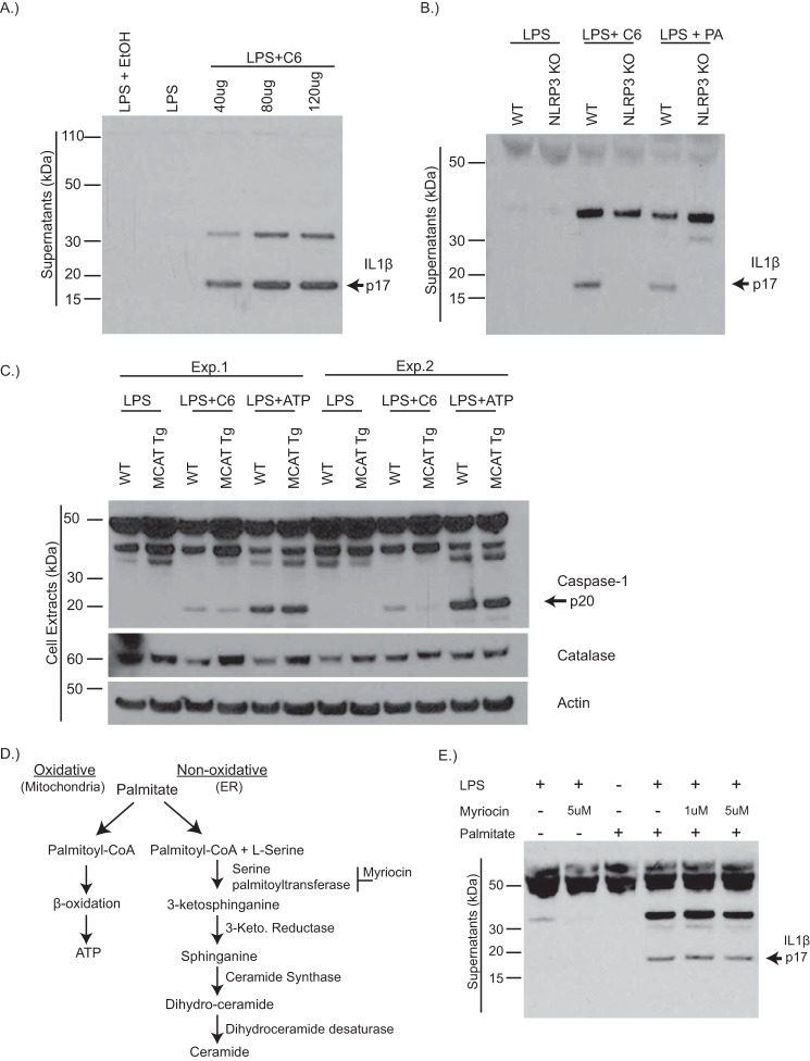
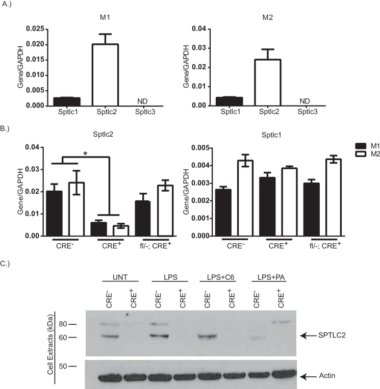
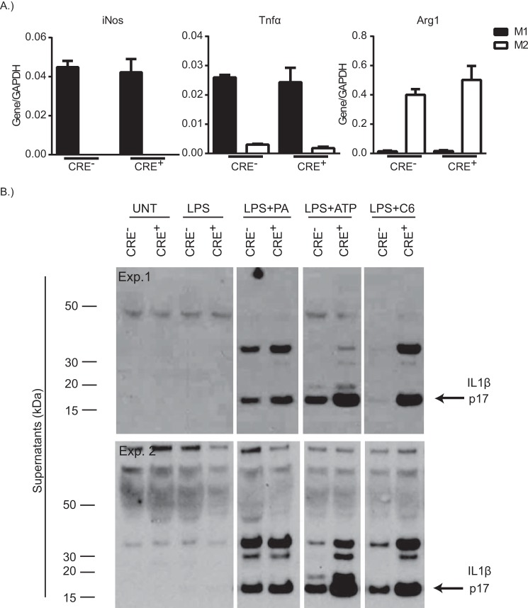
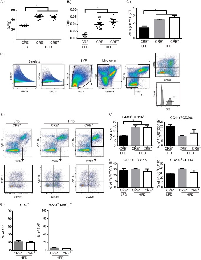
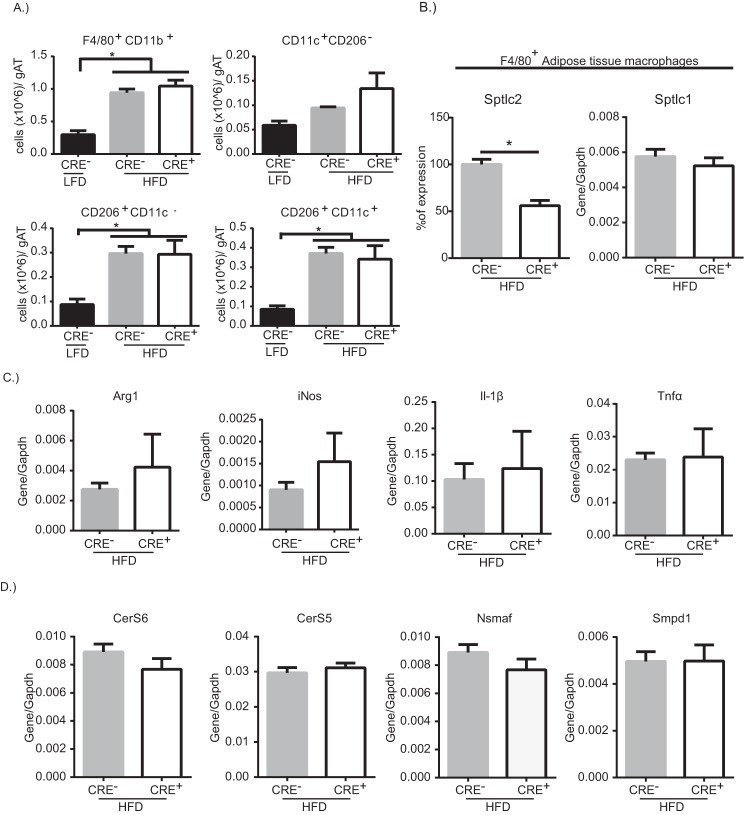
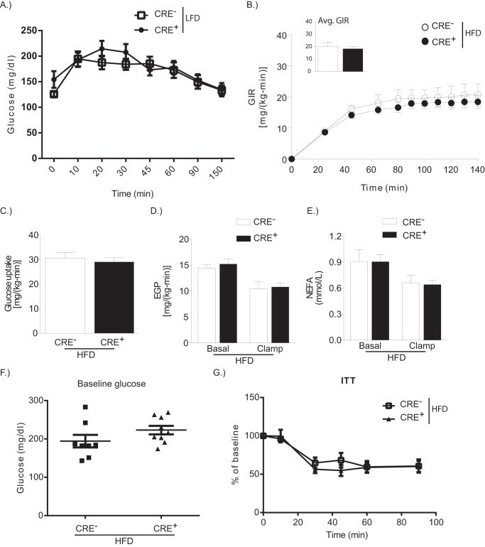
Dietary lipid overload and calorie excess during obesity is a low grade chronic inflammatory state with diminished ability to appropriately metabolize glucose or lipids. Macrophages are critical in maintaining adipose tissue homeostasis, in part by regulating lipid metabolism, energy homeostasis, and tissue remodeling. During high fat diet-induced obesity, macrophages are activated by lipid derived "danger signals" such as ceramides and palmitate and promote the adipose tissue inflammation in an Nlrp3 inflammasome-dependent manner. Given that the metabolic fate of fatty acids in macrophages is not entirely elucidated, we have hypothesized that de novo synthesis of ceramide, through the rate-limiting enzyme serine palmitoyltransferase long chain (Sptlc)-2, is required for saturated fatty acid-driven Nlrp3 inflammasome activation in macrophages. Here we report that mitochondrial targeted overexpression of catalase, which is established to mitigate oxidative stress, controls ceramide-induced Nlrp3 inflammasome activation but does not affect the ATP-mediated caspase-1 cleavage. Surprisingly, myeloid cell-specific deletion of Sptlc2 is not required for palmitate-driven Nlrp3 inflammasome activation. Furthermore, the ablation of Sptlc2 in macrophages did not impact macrophage polarization or obesity-induced adipose tissue leukocytosis. Consistent with these data, investigation of insulin resistance using hyperinsulinemic-euglycemic clamps revealed no significant differences in obese mice lacking ceramide de novo synthesis machinery in macrophages. These data suggest that alternate metabolic pathways control fatty acid-derived ceramide synthesis in macrophage and the Nlrp3 inflammasome activation in obesity.

Keywords: adipose tissue; ceramide synthesis; inflammasome; insulin resistance; macrophage; obesity; saturated fat.


Figures

FIGURE 1.
FIGURE 1.
Ceramides activate the Nlrp3 inflammasome via mitochondrial oxidative stress. A, representative Western blot analysis of IL1β (active p17) protein in supernatants of WT BMDMs that have been primed with LPS for 4 h and stimulated with increasing doses of ceramide (C6) for 6 h. B, Western blot analysis of IL1β protein in BMDMs from WT or Nlrp3−/− mice that have been stimulated with LPS, LPS plus C6 (80 μg/ml) for 6 h, or LPS plus palmitate (400uM) for 24 h. C, Western blot analysis of caspase-1 (active p20) and catalase protein in BMDMs from WT or MCAT transgenic mice that have been treated with LPS alone, LPS plus C6, or LPS plus ATP (5 mm) for 1 h. Two representative blots from individual experiments are shown. D, schematic depicting palmitate entry into oxidative pathway to generate ATP or the nonoxidative pathway causing the de novo synthesis of ceramide. E, Western blot analysis of IL1β protein in the supernatants of WT BMDMs, treated with LPS or LPS plus palmitate, in which some received pretreatment with serine palmitoyltransferase inhibitor, myriocin. The data are representative of two or three individual experiments. Exp., experiment.
FIGURE 2.
FIGURE 2.
Myeloid cell-specific deletion of Sptlc2. A, gene expression of Sptlc1, Sptlc2, and Sptlc3, normalized to gapdh, in WT BMDMs that have been polarized to M1 (LPS (1 μg/ml) plus IFNγ (20 ng/ml)) or M2 (IL4 (10 ng/ml)). B, gene expression of Sptlc2 or Sptlc1 normalized to gapdh in M1 (filled bars) or M2 (open bars) polarized BMDMs from CRE, CRE+, or fl/−; CRE+. C, Western blot analysis of SPTLC2 protein expression in treated BMDMs from CRE or CRE+ mice. Actin is shown as a loading control (n = 2–3 individual experiments; t test). *, p < 0.05. The error bars represent means ± S.E.
FIGURE 3.
FIGURE 3.
Sptlc2 is not required for macrophage polarization or inflammasome activation in vitro. A, iNos, Tnfα, or arg1 gene expression, normalized to gapdh, from CRE or CRE+ BMDMs that have been polarized to M1 (filled bars) or M2 (open bars). B, two Western blots from individual experiments of IL1β protein in supernatants of CRE or CRE+ BMDMs following no treatment, treatment with LPS alone, LPS plus palmitate, LPS plus ATP, or LPS plus C6 (n = 2–3 individual experiments). The error bars represent means ± S.E. Exp., experiment.
FIGURE 4.
FIGURE 4.
Myeloid cell-specific Sptlc2 is not required for diet-induced inflammation. A–C, Body weight (BW, A), visceral adipose tissue weight (AT, B), and cells per gram of adipose tissue (gAT, C) after 13 weeks of LFD or HFD in CRE or CRE+ mice. D, gating strategy to analyze stromavascular fraction (SVF) of adipose tissue. E, representative dot plots of F4/80+CD11b+ cells from adipose tissue of CRE mice on LFD, CRE mice on HFD, or CRE+ mice on HFD. F4/80+CD11b+ cells were gated on to analyze CD206 and CD11c expression. F, quantification of the percentage of macrophage and the macrophage subpopulations. G, quantification of the percentage of lymphocytes, CD3+ T cells, and B220+ B cells, in adipose tissue from HFD-mice (n = 9–10 biological replicates; one-way ANOVA or t test as appropriate). *, p < 0.05. The error bars represent means ± S.E.
FIGURE 5.
FIGURE 5.
Sptlc2-deficient adipose tissue macrophages maintain HFD-induced inflammation. A, quantification of the cells/g AT for macrophages and the macrophage subpopulations from the visceral adipose tissue of CRE or CRE+ mice on HFD. B, expression level of Sptlc2 or Sptlc1 in isolated F4/80+ adipose tissue macrophages from CRE or CRE+ mice on HFD. C and D, gene expression of arg1, iNos, and Il1β Tnfα (C) and CerS6, CerS5, Nsmaf, and Smpd1 (D) (normalized to Gapdh) in isolated adipose tissue macrophages (n = 9–10 biological replicates; one-way ANOVA or t test as appropriate). *, p < 0.05. The error bars represent means ± S.E. gAT, gram of adipose tissue.
FIGURE 6.
FIGURE 6.
Macrophage-specific Sptlc2 is not required for diet-induced insulin resistance. A, glucose tolerance test on LFD-fed CRE or CRE+ mice. B, glucose infusion rates (GIR) during hyperinsulinemic-euglycemic clamps in CRE or CRE+ mice on HFD for 12 weeks. The inset shows the average glucose infusion rate (Avg. GIR). C, whole body glucose uptake. D, endogenous glucose production (EGP) at basal and after clamp. E, nonesterified fatty acid level in the blood at basal and after clamp (n = 4 CRE; n = 7 CRE+). F and G, baseline glucose levels after a 4-h fast (F) and at time points after intraperitoneal injection of insulin (G) (insulin tolerance test, ITT) in CRE or CRE+ mice on HFD (n = 8; t test). *, p < 0.05. The error bars represent means ± S.E.

References

    1. Hossain P., Kawar B., and El Nahas M. (2007) Obesity and diabetes in the developing world: a growing challenge. N. Engl. J. Med. 356, 213–215 - PubMed
    1. Gregor M. F., and Hotamisligil G. S. (2011) Inflammatory mechanisms in obesity. Annu. Rev. Immunol. 29, 415–445 - PubMed
    1. McNelis J. C., and Olefsky J. M. (2014) Macrophages, immunity, and metabolic disease. Immunity 41, 36–48 - PubMed
    1. Lumeng C. N., and Saltiel A. R. (2011) Inflammatory links between obesity and metabolic disease. J. Clin. Invest. 121, 2111–2117 - PMC - PubMed
    1. Nishimura S., Manabe I., Nagasaki M., Eto K., Yamashita H., Ohsugi M., Otsu M., Hara K., Ueki K., Sugiura S., Yoshimura K., Kadowaki T., and Nagai R. (2009) CD8+ effector T cells contribute to macrophage recruitment and adipose tissue inflammation in obesity. Nat. Med. 15, 914–920 - PubMed

Publication types

MeSH terms

LinkOut - more resources