. 2015 Oct 20;112(42):13051-6.
doi: 10.1073/pnas.1517201112. Epub 2015 Oct 5.

cKit+ cardiac progenitors of neural crest origin

Affiliations

cKit+ cardiac progenitors of neural crest origin

Konstantinos E Hatzistergos et al. Proc Natl Acad Sci U S A. .

Abstract

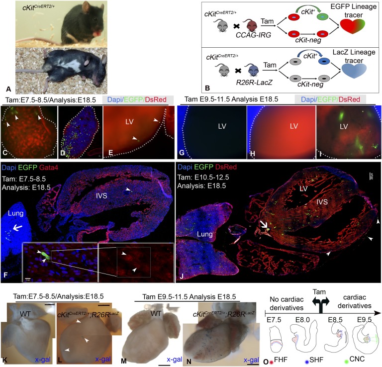
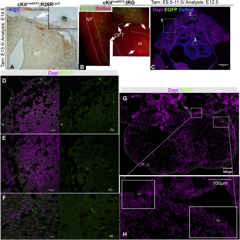
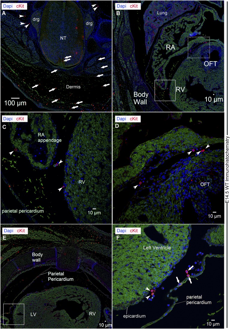
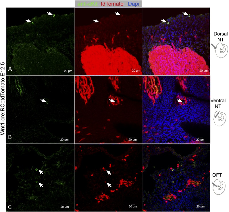
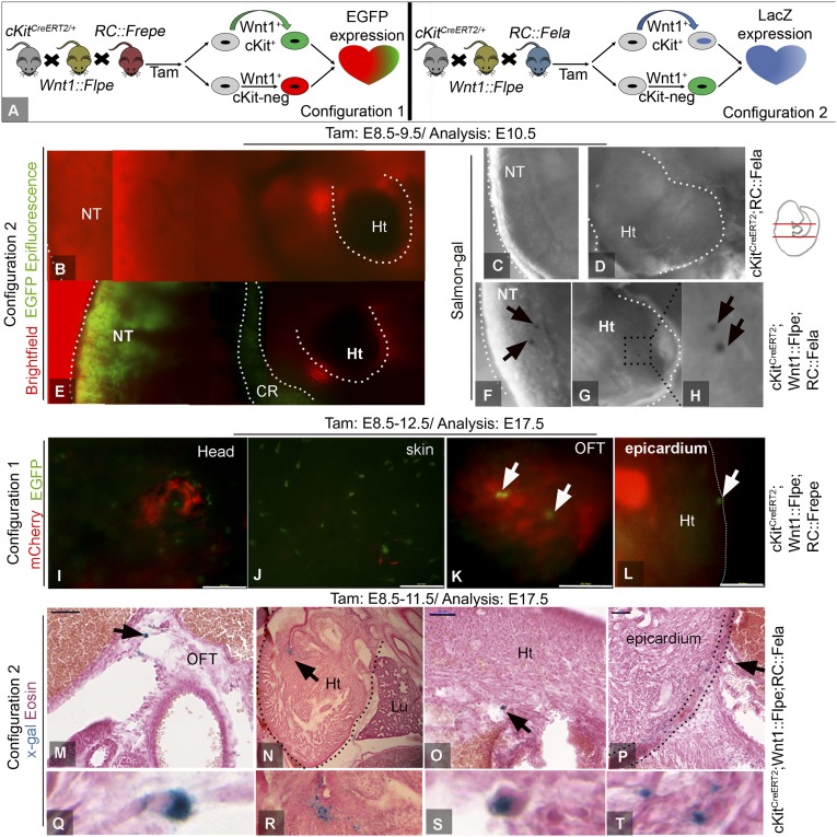
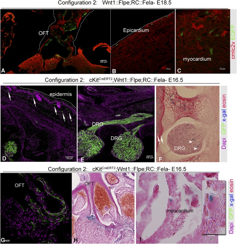
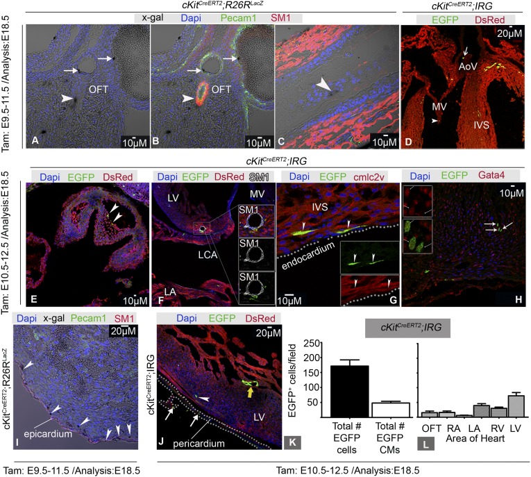
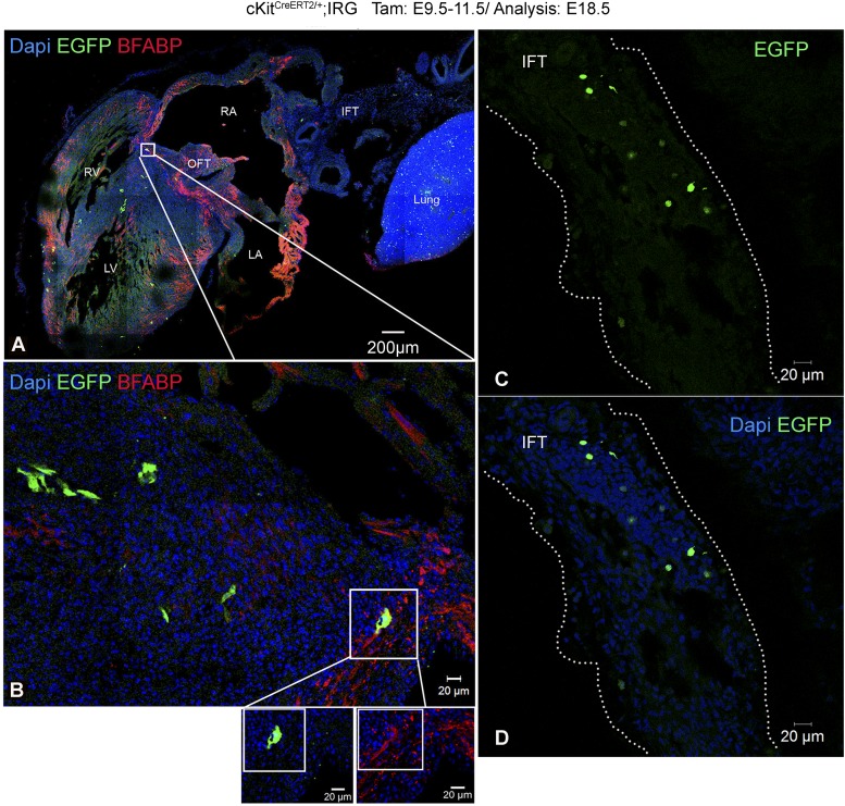
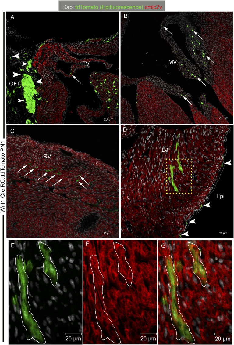
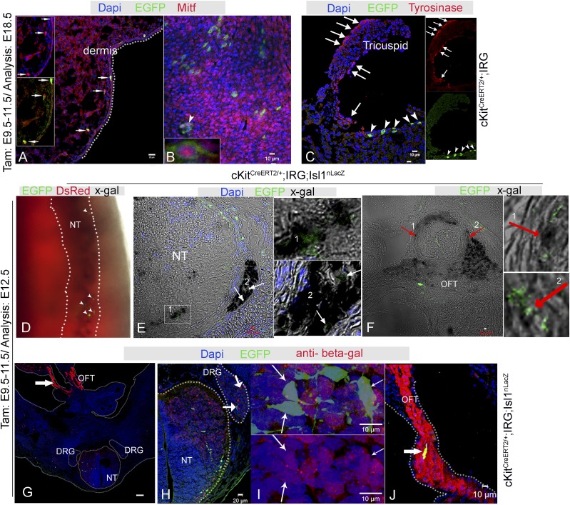
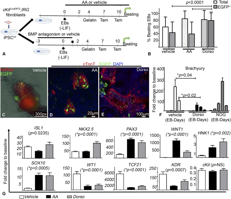
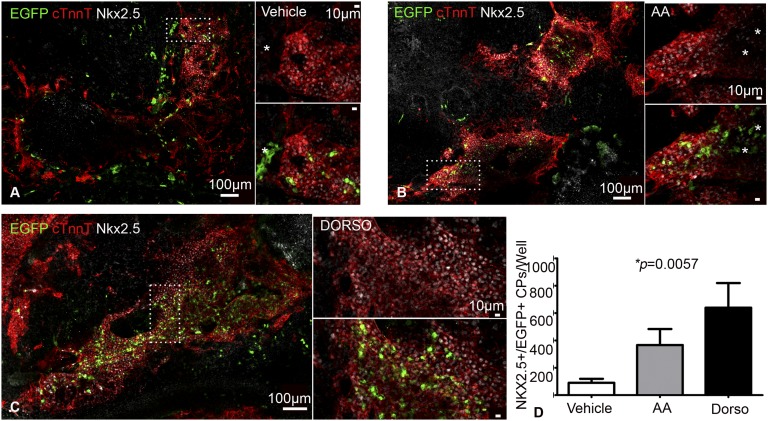
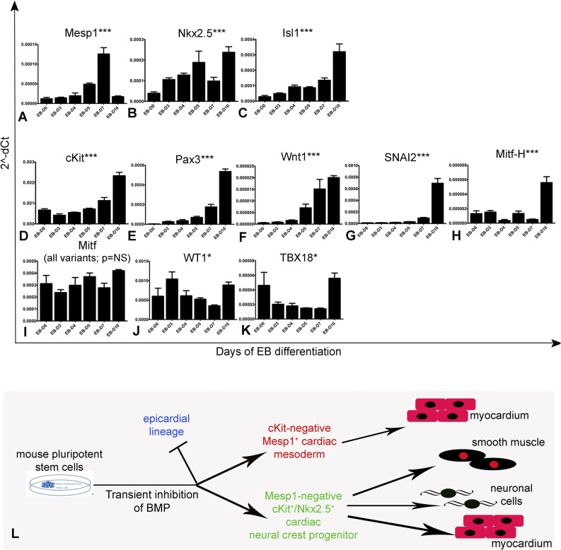
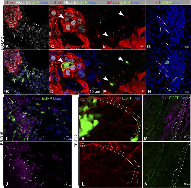
The degree to which cKit-expressing progenitors generate cardiomyocytes in the heart is controversial. Genetic fate-mapping studies suggest minimal contribution; however, whether or not minimal contribution reflects minimal cardiomyogenic capacity is unclear because the embryonic origin and role in cardiogenesis of these progenitors remain elusive. Using high-resolution genetic fate-mapping approaches with cKit(CreERT2/+) and Wnt1::Flpe mouse lines, we show that cKit delineates cardiac neural crest progenitors (CNC(kit)). CNC(kit) possess full cardiomyogenic capacity and contribute to all CNC derivatives, including cardiac conduction system cells. Furthermore, by modeling cardiogenesis in cKit(CreERT2)-induced pluripotent stem cells, we show that, paradoxically, the cardiogenic fate of CNC(kit) is regulated by bone morphogenetic protein antagonism, a signaling pathway activated transiently during establishment of the cardiac crescent, and extinguished from the heart before CNC invasion. Together, these findings elucidate the origin of cKit(+) cardiac progenitors and suggest that a nonpermissive cardiac milieu, rather than minimal cardiomyogenic capacity, controls the degree of CNC(kit) contribution to myocardium.

Keywords: BMP signalling; cardiac neural crest; cardiac stem cells; cardiomyogenesis.


Conflict of interest statement

Conflict of interest statement: K.E.H. and J.M.H. report having a patent for cardiac cell-based therapy. K.E.H. and J.M.H. own equity in Vestion Inc. and are members of the scientific advisory board and consultants of Vestion, Inc. J.M.H. is a board member of Vestion Inc. Vestion Inc. did not participate in funding this work. The other authors report no conflicts.

Figures

Fig. 1.
Fig. 1.
cKitCreERT2/+ lineage-tracing. (A) Phenotype of cKitCreERT2/+ mice. (B) Summary of the experimental design. (C–F) administration of TAM during E7.5–E8.5 (n = 10) marks testicular (C, arrowheads), pulmonary (D) and, rarely, immature cells in the myocardium (E and F, arrowheads). (G–J) Live tissue imaging of cKitCreERT2/+ (G), IRG (H), and cKitCreERT2;IRG (I) E18.5 littermates subjected to TAM during E9.5–E11.5 (n = 7). Widespread EGFP epifluorescence in ventricles and atria (I and J), lungs (J), OFT (J, arrow), epicardium (J, arrowheads). (K–N) Lineage-tracing in cKitCreERT2;R26RlacZ mice (n = 8). (O) Summary of cKit genetic fate-mapping. Panels F and J are confocal tile-scans. Panels KN are photomerged image tiles. (Scale bars, 10 µm in D and F; 200 µm in J; 500px in K–N.) (Magnification, 100× in C, E, and GI.)
Fig. S1.
Fig. S1.
cKit is not expressed in the embryonic myocardium at the time of TAM administration. (A) Colocalization of X-gal with anti-cKit immunohistochemistry (arrow) in an E13.5 NT of a cKitCreERT2/R26RlacZ embryo. (Magnification, 200×.) Inset depicts a higher-magnification image of the indicated cell. (B) Live embryo imaging of EGFP and DsRed epifluorescence in an E12.5 cKitCreERT2;IRG embryo. Two EGFP+ cells are detected in proximity to the OFT and two more in the epicardium (arrows). EGFP expression is absent in the myocardium. In contrast, strong expression of EGFP is seen in the NT and the skin. (C–F) A transverse section from an E12.5 cKitCreERT2;IRG mouse embryo in which immunohistochemistry against EGFP has been performed. EGFP cells are detected in the skin (Inset 1 and D in higher magnification), neural tube (Inset 2 and E in higher magnification), and the conotruncus (Inset 3 and F in higher magnification). No EGFP signal is detected in the myocardium. (G and H) A transverse section of an E12.5 cKitCreERT2;IRG heart illustrating expression of EGFP in the epicardium and left atrium. No signal is detected in the myocardium. Panel B is a photomerged image tile. (Magnification, 100×/tile.) Panel C is a confocal tile-scan. Ht, Heart. (Scale bars, 10 µm in D–F, and 100 µm in GH.)
Fig. S2.
Fig. S2.
Expression of cKit in the cKit+/+ developing mouse heart. Representative confocal immunofluorescence of cKit antibody localization in E14.5 wild-type mouse embryos. (A) Consistent with cKitCreERT2/+ genetic fate map, CNCkit (red fluorescence) are detected in the NT, DRG, as well as in the dermis. CNCkit are detected in proximity of both the dorsolateral (arrows) and ventral (arrowheads) sites of the NT. (B) cKit expression in the embryonic lungs, RA, OFT, and epicardium (boxes). (C) Magnification of the solid box in B, highlights CNCkit (arrowheads) in the RA appendage, pericardial and epicardial walls. (D) Magnification of the dashed box in B, highlights CNCkit (arrowheads) accessing the RA, epicardium, and OFT. (E) CNCkit are dispersed in dermis, parietal pericardium, epicardium, and cardiac ventricular walls. (F) Magnification of the boxed area in E, illustrates CNCkit accessing the LV through the pericardial (arrows) and epicardial (arrowheads) walls. (Scale bars, 100 µm in A and 10 µm in B–F.) RA, right atrium; RV, right ventricle.
Fig. S3.
Fig. S3.
cKit immunohistochemistry labels weakly expressing tdTomato+ cells in Wnt1-Cre;RC::tdTomato mouse embryos. Confocal immunofluorescence analysis following anti-cKit immunohistochemistry in E12.5 Wnt1-Cre;RC::tdTomato embryos illustrates colocalization of cKit in a population of cells with weak tdTomato epifluorescence, located dorsally (A, arrows) and ventrally from the NT (B, arrow), as well as within the outflow tract (C, arrows). A total of n = 3 Wnt1-Cre;RC::tdTomato embryos were analyzed. (Scale bars, 20 µm.)
Fig. 2.
Fig. 2.
Intersectional genetic fate-mapping of cKit and Wnt1. (A) schematic of the two different approaches of the study. (B–D) Live epifluorescence imaging (B), followed by salmon-gal histochemical detection of nLacZ (C and D), in an E10.5 cKitCreERT2;RC::Fela embryo exposed to TAM during E8.5–E9.5. The flp- and intersectional indicators are not expressed in the absence of Flpe and Flpe/Cre-mediated recombination, respectively. (E–H) A Wnt1::Flpe;cKitCreERT2;RC::Fela littermate exhibits widespread GFP epifluorescence (E) and a few salmon-gal+ cells in the NT and heart. (I–L) Live embryo imaging of mCherry and GFP epifluorescence in a E17.5 Wnt1::Flpe;cKitCreERT2;RC::Frepe embryo. EGFP+ CNCkit in the craniofacial region (I), skin (J), OFT (K), and the epicardial wall of the heart (L). (M and T) X-gal+ CNCkit derivatives in the OFT (M and G), heart (N, O, R, and S), and epicardium (P and T) of E17.5 Wnt1::Flpe;cKitCreERT2;RC::Fela embryos. Arrows in M–P are depicted in higher magnification in panels Q–T, respectively. Panels B, E are photomerged image tiles. OFT, outflow tract; Ht, heart; Lu, lung. (Scale bars, 50 µm in M–P and 300px in I–L.) (Magnification, 100× in BH and QT.)
Fig. S4.
Fig. S4.
Intersectional genetic fate map of cKit and Wnt1. A-C, Wnt1::Flpe4351 reliably marks the cardiac neural crest as indicated by the expression of EGFP in the OFT (A), epicardium (B), and myocardium (C) of an E18.5 Wnt1::Flpe;Rc::Fela heart. (D–I) When both Wnt1::Flpe and cKitCreERT2/+ become activated in cKitCreERT2;Wnt1::Flpe;Rc::Fela embryos, expression of EGFP is still widely expressed in the NC derivatives, including melanoblasts (D, arrows), DRGs (E), and OFT (G). In addition, a population of nLacZ+ cells are also present within the skin (F, arrows), DRGs (F, arrowheads), OFT (H) and, rarely, within the compact myocardium (I). [Scale bars, 100 µm (A, D, and E), 10 µm (B and C), 20 µm (G) and 80 µm (I).] (Magnification, 100× in H.)
Fig. 3.
Fig. 3.
Heart derivatives of CNCkit. (A and B) Colocalization of X-gal with SM1 (arrowhead) and Pecam1 (arrows) in the OFT. (C) X-gal+ cells (arrowhead) within the aortic tunica media. (D) EGFP+ derivatives within the mitral valve (arrowhead) and aortic valve (arrow). (E) EGFP+ derivatives in the aortic valve (arrowheads). (F) CNCkit are associated with, but do not contribute to, coronary vasculature. (G) EGFP+/cmlc2v+ ventricular cardiac myocytes. (H) EGFP+ cardiac derivatives coexpress Gata4+. (I and J) EGFP+ pericardium (arrows), epicardium (arrowheads), and endocardium (J, yellow arrow). (K) Cardiomyocytic versus noncardiomyocytic EGFP+ derivatives in the heart. (L) Distribution of EGFP+ cells in the heart [n = 3 embryos; 11 sections (K and L)]. AoV, aortic valve; CM, cardiomyocytes; cmlc2v, cardiac muscle light chain 2v; LA, left atrium; LV, left ventricle; LCA, left coronary artery; MV, mitral valve; SM1, smooth muscle myosin heavy chain. Values represent means ± SEM.
Fig. S5.
Fig. S5.
CNCkit derivatives in the heart and their identity. (A) Tilescan image illustrating extensive contribution of EGFP+ CNCkit derivatives in the lung, IFT, atria, ventricles and OFT. Notably, EGFP+ cells are consistently detected to be closely associated with BFABP+ satellite glial progenitors. Occasionally, EGFP and BFABP colocalize, illustrating that CNCkit contribute to glia and glial progenitor lineages (B, boxed area). (C and D) Higher magnification of the IFT from A, illustrating extensive contribution from CNCkit. BFABP, brain fatty acid-binding protein; IFT, inflow tract; LA, left atrium; LV, left ventricle; RA, right atrium; RV, right ventricle.
Fig. S6.
Fig. S6.
Wnt1-expressing CNCs contribute cardiomyocytes in the murine heart. Similar to the CNCkit derivatives, confocal microscopy of tdTomato (epifluorescence; pseudocolored green) in postnatal day 1 (PN1) Wnt1-Cre;RC::tdTomato mouse hearts illustrates that the Wnt1-expressing CNCs contribute extensively in the OFT (A, arrowheads), tricuspid valve (A, arrow), and mitral valve (B, arrows). In addition, tdTomato+ cells are detected within the ventricular myocardium (C, arrows, and D; yellow Inset) and epicardium (D, arrowheads). Immunostaining against the cardiomyocyte-specific marker cmlc2v illustrates that, similar to the CNCkit, the Wnt1-expressing CNCs contribute both noncardiomyocytes (C, arrows; tdTomato+/cmlc2v) and fully differentiated cardiomyocytes (D, Inset; tdTomato+/cmlc2v+) in the mouse ventricle. (E–G) Higher magnification of the tdTomato+/cmlc2v+ clone of ventricular cardiomyocytes in the yellow Inset in D. (Scale bars, 20 µm.)
Fig. S7.
Fig. S7.
Mitf and Isl1 expression in CNCkit. (A) EGFP+ melanoblasts in the skin (arrows) coexpress Mitf. (B) EGFP+ derivatives in the heart (arrowhead, Inset) coexpress Mitf. (C) Tyrosinase+ melanocytes in the tricuspid valve (arrows) do not colocalize with EGFP (arrowheads). (D) Colocalization of X-gal and EGFP epifluorescence in cKitCreERT2;IRG;Isl1nLacZ embryos. (Magnification, 100×.) (E) X-gal and EGFP colocalization in the NT (Inset, 1) and DRGs (arrows, 2). (F) X-gal and EGFP colocalization (1, 2, arrows) in the OFT. (G) Colocalization of EGFP and β-gal in the NT, DRGs, and OFT (arrow). (H) EGFP and β-gal colocalization in NT and DRGs. (I) EGFP+/β-gal+ neurons (arrows) in the NT. (J) Higher magnification of the OFT in (G, arrow). [Scale bars, 10 µm (B, C, E, F, I, J), 100 μm (G), and 20 µm (A and H).]
Fig. 4.
Fig. 4.
Transient BMP antagonism in iPSCkit induces CNCkit and suppresses the epicardium. (A) Schematic of the experimental approach. (B) Quantification of the percentage of beating EBs, and the percentage of beating EBs that are EGFP+ following Cre-recombination. (C) Live fluorescent imaging of a vehicle-treated spontaneously beating EB, coexpressing EGFP and DsRed. (D and E) Confocal microscopy of EBs following treatment with AA (D) or Dorso (E), illustrates that the EGFP+ cKitCreERT2/+ derivatives within the EBs are cTnnT+ cardiomyocytes. (F) Gene-expression analysis of Brachyury during the time-course of iPSCkit differentiation into cardiomyocytes following treatment with vehicle, Dorso, or NOG. (G) Comparison of the expression profiles of cardiac mesoderm- and CNC-related genes in day 11 EBs, in response to treatment with vehicle, AA, or Dorso. Compared with controls, AA and Dorso enhance cardiomyogenesis via a significant induction in CNC-related genes while suppressing proepicardial and endothelial progenitor genes. In addition, Dorso significantly enhances the expression of ISL1 and NKX2.5. cTnnt, cardiac troponin T; values represent means ± SEM.
Fig. 5.
Fig. 5.
Derivation of CNCkit from mouse iPSCs. (A–C) Representative confocal immunofluorescence images illustrating EGFP+/NKX2.5+ derivatives within cTnnT+ EBs of vehicle-treated (A), AA-treated (B), or Dorso-treated (C) iPSCkit. Note that several of the EGFP+ cells in the vehicle- and AA-treated groups are NKX2.5 (asterisks). (Insets) Higher magnification. (D) Quantitation of EGFP+/NKX2.5+ cells between groups (n = 9 per group). Values represent means ± SEM.
Fig. S8.
Fig. S8.
Transient BMP antagonism induces CNCkit and suppresses epicardium. (A–J) NOG promotes the generation of cardiac mesoderm from iPSCkit as indicated by the transient induction of Mesp1 (A) and up-regulation in the expression of Nkx2.5 (B) and Isl1 (C) at ∼EB- day 7. Subsequently, establishment of cardiac mesoderm is followed by the induction of CNC at ∼EB day 10, as indicated by the dramatic up-regulation in the expression of cKit (D), Pax3 (E), Wnt1 (F), SNAI2 (G), and Mitf-H (H). Notably, the noncardiac variants of Mitf (variants 1 and 2) remain unchanged. (J–K) Expression of the proepicardial lineage markers WT1 and TBX18 is significantly repressed before reaching baseline values by EB day 10. (L) Summary of the iPSC-based lineage tracing experiments. Values represent means ± SEM. ***P < 0.0001, *P < 0.05. n = 3 per group.
Fig. S9.
Fig. S9.
Derivation of CNCkit from mouse iPSCs following NOG-mediated BMP antagonism. (A and B) A cluster of iPSCkit-derived EGFP+/Nkx2.5+, undifferentiated (cTnnT) CNCkit (arrows) next to a cluster of non-CNCkit-derived (EGFP) cTnnT+ differentiated cardiomyocytes. (C and D) An iPSCkit-derived CNCkit fully differentiated into cTnnT+/Nkx2.5+ cardiomyocyte (arrowhead). (E and F) Differentiation of iPSCkit-derived CNCkit into SM22a+ smooth muscle cells. (G and H) Colocalization of iPSCkit-derived CNCkit with Isl1. (I and J) iPSCkit-derived CNCkit generate Pax3+ progenitors. (K and L) EGFP+/neurofilament M+ motorneurons within a spontaneously beating EB. (M and N) An EGFP+/Tuj1+ neuron within the beating EBs. n = 3 per group. (Scale bars, 10 µm in A–F and I–N; 20 µm in G and H.)

Comment in

References

    1. Rana MS, Christoffels VM, Moorman AF. A molecular and genetic outline of cardiac morphogenesis. Acta Physiol (Oxf) 2013;207(4):588–615. - PubMed
    1. Witty AD, et al. Generation of the epicardial lineage from human pluripotent stem cells. Nat Biotechnol. 2014;32(10):1026–1035. - PMC - PubMed
    1. Burridge PW, Keller G, Gold JD, Wu JC. Production of de novo cardiomyocytes: Human pluripotent stem cell differentiation and direct reprogramming. Cell Stem Cell. 2012;10(1):16–28. - PMC - PubMed
    1. Yang L, et al. Human cardiovascular progenitor cells develop from a KDR+ embryonic-stem-cell-derived population. Nature. 2008;453(7194):524–528. - PubMed
    1. Jain R, et al. HEART DEVELOPMENT. Integration of Bmp and Wnt signaling by Hopx specifies commitment of cardiomyoblasts. Science. 2015;348(6242):aaa6071. - PMC - PubMed

Publication types

Substances