Review
. 2015 Nov;13(11):663-76.
doi: 10.1038/nrmicro3524. Epub 2015 Oct 6.

Filovirus pathogenesis and immune evasion: insights from Ebola virus and Marburg virus

Affiliations
Review

Filovirus pathogenesis and immune evasion: insights from Ebola virus and Marburg virus

Ilhem Messaoudi et al. Nat Rev Microbiol. 2015 Nov.

Abstract

Ebola viruses and Marburg viruses, members of the filovirus family, are zoonotic pathogens that cause severe disease in people, as highlighted by the latest Ebola virus epidemic in West Africa. Filovirus disease is characterized by uncontrolled virus replication and the activation of host responses that contribute to pathogenesis. Underlying these phenomena is the potent suppression of host innate antiviral responses, particularly the type I interferon response, by viral proteins, which allows high levels of viral replication. In this Review, we describe the mechanisms used by filoviruses to block host innate immunity and discuss the links between immune evasion and filovirus pathogenesis.


Conflict of interest statement

statement The authors declare no competing interests.

Figures

Figure 1
Figure 1. Filovirus genome structure and life cycle
A. The genome of EBOV is depicted. The genome is depicted in the 3′ to 5′ orientation to indicate that the genomic RNA is negative-sense. The genes are named after the proteins encoded by each. They are NP, nucleoprotein; VP35, viral protein 35; VP40, viral protein 40; GP/sGP, glycoprotein/soluble glycoprotein; VP30, viral protein 30; VP24, viral protein 24; L, Large protein (the viral polymerase). Note that Marburg virus does not encode a sGP protein. Filoviruses have unusually long non-coding regions at the 5′ and 3′ end of their mRNAs. The genome regions corresponding to these non-protein coding sequences are not drawn to scale. B. The general life cycle of a filovirus is displayed. First, the filovirus attaches to the cell membrane via its surface GP protein. Virus is taken up by a process known as macropinocytosis,–. Upon acidification of the endosome, cathepsins B and L (cellular proteases) cleave GP. This allows GP to interact with host protein NPC-1, an interaction which is a prerequisite for fusion of viral and endosomal membranes,. Host endosomal calcium channels called two-pore channels (TPCs) play a critical role in the endosomal trafficking of incoming viral particles to the site of fusion. GP mediates fusion of the viral and the endosomal membrane, releasing the viral ribonucleocapsid into the cytoplasm where the negative strand RNA genome undergoes transcription and replication. Production of 5′-capped, 3′-polyadenylated mRNAs from individual viral genes occurs and genome replication follows, in which the genomic RNA template is copied into a full-length complement. The full-length complement serves as template for synthesis of additional negative-sense genome. The RNA synthesis reactions require the NP, VP35 and L proteins. Initiation of transcription for Ebola virus also requires the VP30 protein, whereas the replication reactions do not require VP30. Viral proteins, including NP, VP35, VP40, GP, VP30, VP24, and L are translated from the viral mRNAs. New viral particles are assembled at the plasma membrane. The VP40 protein functions as the viral matrix protein and directs budding of particles from the cell surface. GP, a type I transmembrane protein, is incorporated into the budding particles, as are viral nucleocapsids containing the viral genome.
Figure 2
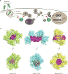
Figure 2. Subversion of IFN induction
A. Schematic of the IFN production pathway and mechanisms of filoviral evasion. The image depicts how EBOV VP35 (eVP35) and MARV VP35 (mVP35) proteins antagonize signaling pathways that lead to IFN-α/β gene expression. RIG-I-like receptors (RLRs) which include RIG-I and MDA5, detect products of viral replication, RNAs such as cytoplasmic dsRNAs or RNAs with 5′-triphosphates. Activation of RLRs is facilitated by the protein PACT. Upon activation, RLRs signal through the mitochondria-associated protein MAVS to activate kinases IKKε and TBK1. These phosphorylate interferon regulatory factor (IRF)-3 or 7 then accumulate in the nucleus and promote IFN-α/β gene expression. VP35 proteins can bind to dsRNAs and to PACT, preventing RLR activation. In addition, VP35s interact with and act as decoy substrates for the kinases IKKε and TBK1. B. Structural analysis of filoviral VP35 protein interferon inhibitory domain (IID) binding to dsRNAs reveals differences among the dsRNA recognition mode for EBOV, RESTV, and MARV VP35 proteins. (Left) The structure of eVP35 bound to 8 bp dsRNA (PDB 3L25). (Middle) rVP35 bound to 12bp (PDB 4LG2). (Right) mVP35 IID bound to 12 bp dsRNA (PDB 4GHL). dsRNA shown in magenta.
Figure 3
Figure 3. Subversion of IFN-induced signaling and of IFN-induced protein tetherin
A. Schematic of the IFN response pathway and mechanisms of filoviral evasion. IFN-α/β binds the extracellular domains of the heterodimeric IFN alpha receptor composed of subunits IFNAR1 and IFNAR2. This activates receptor-associated tyrosine kinases Jak1 and Tyk2. These phosphorylate STAT1 and STAT2 causing them to heterodimerize, associate with karyopherin alpha (KPNA)-1, 5 and 6 and be transported into the nucleus to induce interferon stimulating genes, including the antiviral kinase PKR and major histocompatibility complex 1 MARV VP40 (mVP40) inhibits the IFN induced activation of tyrosine kinase JAK1. EBOV VP24 (eVP24) binds KPNA1, 5 and 6 to block KPNA-STAT1 interactions. Both mechanisms inhibit IFN-induced gene expression. B. EBOV VP24 uses a unique non-classical nuclear localization site to interact with KPNA-1, 5 or 6. (Left) EBOV VP24 (PDB 4M0Q) and SUDV VP24 (PDB 3VNF) show a similar pyramidal structure. (Right) eVP24 proteins bind the KPNA C-terminus (PDB 4U2X). C. The IFN-inducible protein tetherin can restrict budding of the filovirus VP40 protein (left). However, filovirus GP counteracts the antiviral effects of tetherin, allowing Ebola and Marburg viruses to be released in the present of tetherin (right).
Figure 4
Figure 4. Subversion of host translation mechanisms to support virus replication
It has been observed that several of the 5′-capped viral EBOV mRNAs possess upstream open reading frames (uORFs) in their 5′ untranslated regions (UTRs). Under lowstress conditions, the uORF present in the mRNA encoding the L protein attenuates L translation initiation. This is because scanning ribosomes (depicted as ovals) initiate efficiently at the uORF AUG instead. However, under stress, such as occurs due to activation of innate immune responses, eIF2α phosphorylation (eIF2α~P) increases and translation initiation at the uORF becomes less efficient. This allows the scanning ribosome to bypass the uORF AUG and increases initiation at the L AUG, helping to sustain L expression despite the increase in eIF2α phosphorylation.

References

    1. Baize S, et al. Emergence of Zaire Ebola Virus Disease in Guinea – Preliminary Report. N Engl J Med. 2014;371:1418–1425. doi: 10.1056/NEJMoa1404505. - DOI - PubMed
    1. Feldmann H, Geisbert TW. Ebola haemorrhagic fever. Lancet. 2010;377:849–862. doi: 10.1016/S0140-6736(10)60667-8. - DOI - PMC - PubMed
    1. Sanchez A, Geisbert TW, Feldmann H. In: Fields Virology. Knipe DM, Howley PM, et al., editors. Lippincott Williams and Wilkins; 2007. pp. 1410–1448.
    1. Gire SK, et al. Genomic surveillance elucidates Ebola virus origin and transmission during the 2014 outbreak. Science. 2014;345:1369–1372. - PMC - PubMed
    1. WHO. Ebola Situation Report – 4 February 2015. 2015

Publication types

MeSH terms

Substances