Review
. 2015 Nov;23(11):693-706.
doi: 10.1016/j.tim.2015.07.010. Epub 2015 Oct 1.

Bacterial amyloid formation: structural insights into curli biogensis

Affiliations
Review

Bacterial amyloid formation: structural insights into curli biogensis

Nani Van Gerven et al. Trends Microbiol. 2015 Nov.

Abstract

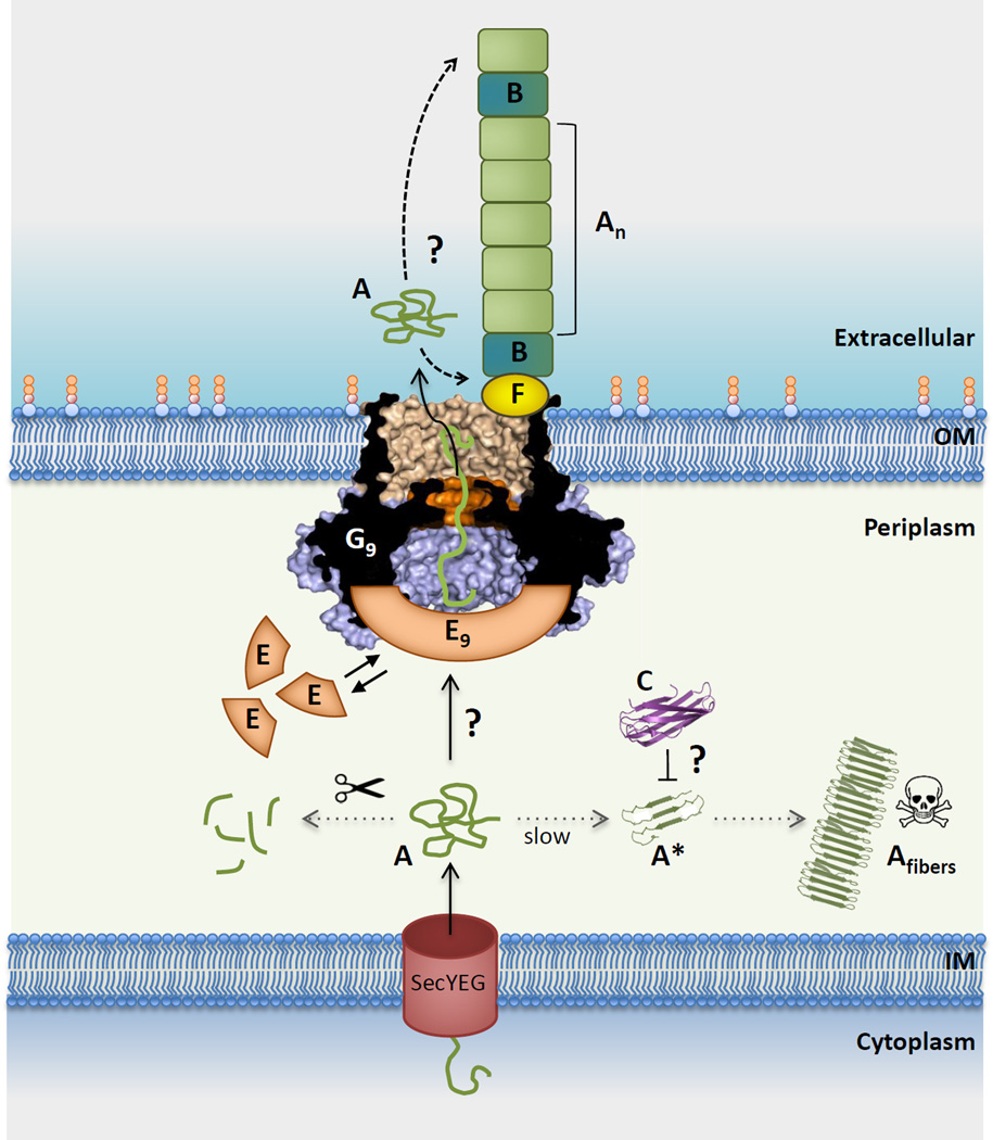
Curli are functional amyloid fibers assembled by many Gram-negative bacteria as part of an extracellular matrix that encapsulates the bacteria within a biofilm. A multicomponent secretion system ensures the safe transport of the aggregation-prone curli subunits across the periplasm and outer membrane, and coordinates subunit self-assembly into surface-attached fibers. To avoid the build-up of potentially toxic intracellular protein aggregates, the timing and location of the interactions of the different curli proteins are of paramount importance. Here we review the structural and molecular biology of curli biogenesis, with a focus on the recent breakthroughs in our understanding of subunit chaperoning and secretion. The mechanistic insight into the curli assembly pathway will provide tools for new biotechnological applications and inform the design of targeted inhibitors of amyloid polymerization and biofilm formation.

Keywords: amyloid chaperone; biofilm matrix; functional amyloid; peptide diffusion channel; protein secretion.


Figures

Figure 1
Figure 1. Curli composition and structure
(A) Organization of the csgBAC and csgDEFG curli operons and architecture of the curli subunits CsgA (light green) and CsgB (dark green). The N-terminal signal sequence (SEC; red) is cleaved after export into the periplasm. The mature subunits contain an N-terminal curli-specific targeting sequence (N22 or N23 in CsgA and CsgB, respectively) that is followed by a pseudo-repeat region (R1 to R5) that forms the amyloidogenic core of the curli subunits (green). Repeats that efficiently self-polymerize in vitro are underscored. (B) Electron microscopy of curli fibers. (i, ii) Freeze-fracture EM of E. coli biofilms shows bacterial cells are encased in a matrix supported by interwoven curli. Bacteria appear to come into contact with the matrix only at discrete locations (white arrows); (m: fractioned bacterial membrane); scale bars 500 nm. (reproduced from [12]). (iii, iv) Transmission EM of individual E. coli cells producing curli fibers (iii), and curli-like fibers grown in vitro from purified CsgA (iv); scale bars: 200 nm. (C) Representation of typical in vitro CsgA polymerization profiles under different conditions. The addition of preformed fibers or the CsgB nucleator removes the lag phase preceding exponential fiber growth (blue curve). In the presence of CsgE (1:1 ratio) or CsgC (1:500 ratio), no CsgA polymerization is observed (black curve) [24].
Figure 2
Figure 2. Architecture and 3D model of curli subunit CsgA
(A) CsgA amino acid sequence aligned to highlight the conserved motifs (black boxes) in N22 and the pseudo-repeat structure in R1 to R5. Residues forming the predicted inner core of the β-helix in the modeled CsgA structures are differentiated in color. Indicated in red squares are gatekeeper residues that reduce aggregation propensity of individual repeats [85]. (B) Structural models of CsgA. Lowest-energy conformations of theoretical CsgA models predicted based on amino acid covariation analysis [64]. The MD simulations and covariance restraints were compatible with both left-handed (left) and right-handed (right) β-helical structures. For both, the width of the β-helical core is around 31 Å. The ’rectangular’ hydrophobic cores are primarily formed by side chains of Ala, Ile, Leu, Met and Val, and colored as in (A). Future experimental characterization is required to validate whether either model is representative of the CsgA structure in the curli fibers.
Figure 3
Figure 3. Structure and function of the curli chaperone CsgC
(A) Cartoon representation of the X-ray structure of oxidized CsgC (PDB 2Y2Y; [67]). The disulfide bond connecting C29 to C31 is shown in orange. (B) Schematic representation of proposed role of CsgC in CsgA chaperoning, showing a predicted energy landscape of CsgA amyloidogenesis where CsgA forms dynamic, amorphous aggregates before assembling into amyloid-like, prefibrillar oligomers. CsgC is believed to avoid periplasmic CsgA amyloidosis by transiently interacting with a prefibrillar CsgA and inhibiting its progression as a template for CsgA fibrillation. Figure adapted from Evans et al. [24].
Figure 4
Figure 4. The structure of curli secretion channel CsgG
(A, B) Side and cross-sectional view of CsgG nonamers in ribbon and surface representation, respectively. (PDB: 4UV3; [23]) (A) A single protomer is colored from N- to C-terminus in a gradient from blue to red, respectively. The N-terminal lipidation sites are marked by magenta spheres. (B) Helix 2, the core domain and transmembrane (TM) β-hairpins are shown in blue, light blue and tan, respectively. Helix 2 forms a docking side for a CsgE nonamer (panel D) Abbreviations: OM, outer membrane. (C) Close-up view of the constriction loop of a single CsgG protomer. The channel constriction (boxed region in panel B) is formed of stacked rings F56, N55 and Y51; arrow indicates direction of CsgA transport. H-clamp: conserved H-bond donor/acceptor. (D) Top view of the CsgG channel constriction, with a modelled polyalanine chain residing in the channel, viewed form the extracellular side. (E) 3D cryo-EM reconstruction of the CsgG:CsgE complex. The model shows a nonameric particle comprised of CsgG (blue) and an additional density assigned as a CsgE nonamer (orange), encapsulating a pre-constriction chamber of approximately 24.000 Å3. Figures adapted from Goyal et al. [23].
Figure 5
Figure 5. Integrated model for curli subunit secretion
CsgA (A) subunits enter the periplasm via the SecYEG translocon, from where they progress to the cell surface via the curli transporter CsgG (G) or are proteolytically degraded (left dotted line). Premature polymerization of CsgA in the periplasm (right dotted line) is subverted by CsgC (C), probably by the binding and neutralization of early assembly intermediates (labeled A*). CsgG forms a nonameric complex (G9) that acts as a non-specific peptide diffusion channel. A nonameric CsgE complex (E9) acts as specificity and secretion factor to the CsgG channel and forms a capping structure to a pre-constriction cavity in the CsgG complex. Recruitment and (partial) enclosure of CsgA in the pre-constriction cavity is proposed to create an entropy gradient over the channel that favors CsgA’s outward diffusion as an unfolded, soluble polypeptide. Once secreted, curli fiber formation and elongation is templated by CsgB (B), in a CsgF (F)-dependent manner. It is unknown whether fibers grow from the proximal or distal end (dashed arrows). Also, the sequence that leads to the assembly of the CsgF:CsgG:CsgE:CsgA secretion complex, or its stoichiometry (i.e. G9:E9 + n CsgA subunits) are currently unknown [23]), with three possible scenarios as follows: (1) does an encounter of CsgE and CsgA in the periplasm lead to their docking onto CsgG?; (2) or does CsgG itself recruit CsgA prior to the binding of the capping CsgE nonamer?; (3) or are CsgG and CsgE in a constitutive complex that is dynamically gated in function of CsgA binding? Further experimental and structural work is needed to resolve these mechanistic questions in the curli secretion pathway. Abbreviations: IM, inner membrane; OM, outer membrane.

References

    1. Hall-Stoodley L, Stoodley P. Evolving concepts in biofilm infections. Cell. Microbiol. 2009;11:1034–1043. - PubMed
    1. Branda SS, et al. Biofilms: the matrix revisited. Trends Microbiol. 2005;13:20–26. - PubMed
    1. Anderson GG, O’Toole GA. Innate and induced resistance mechanisms of bacterial biofilms. Curr. Top. Microbiol. Immunol. 2008;322:85–105. - PubMed
    1. Høiby N, et al. Antibiotic resistance of bacterial biofilms. Int. J. Antimicrob. Agents. 2010;35:322–332. - PubMed
    1. Gilbert P, et al. The physiology and collective recalcitrance of microbial biofilm communities. Adv. Microb. Physiol. 2002;46:202–256. - PubMed

Publication types

MeSH terms