Case Reports
. 2015 Oct 6:5:14829.
doi: 10.1038/srep14829.

Molecular investigation of coexistent chronic myeloid leukaemia and peripheral T-cell lymphoma - a case report

Affiliations
Case Reports

Molecular investigation of coexistent chronic myeloid leukaemia and peripheral T-cell lymphoma - a case report

Alicja M Gruszka et al. Sci Rep. .

Abstract

Chronic myeloid leukemia (CML) is a myeloproliferative neoplasm underlain by the formation of BCR-ABL1 - an aberrant tyrosine kinase - in the leukaemic blasts. Long-term survival rates in CML prior to the advent of tyrosine kinase inhibitors (TKIs) were dismal, albeit the incidence of secondary malignancies was higher than that of age-matched population. Current figures confirm the safety of TKIs with conflicting data concerning the increased risk of secondary tumours. We postulate that care has to be taken when distinguishing between coexisting, secondary-to-treatment and second in sequence, but independent tumourigenic events, in order to achieve an unbiased picture of the adverse effects of novel treatments. To illustrate this point, we present a case of a patient in which CML and peripheral T-cell lymphoma (PTCL) coexisted, although the clinical presentation of the latter followed the achievement of major molecular response of CML to TKIs.


Figures
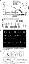
Figure 1
Figure 1
(A) Clinical and treatment history of the patient. Hb – haemoglobin, PLT – platelets, WBC – white blood count, HU – hydroxyurea, CHOP - cyclophosmamide, hydroxydaunorubicin, oncovin (vincristine), prednisone, IGEV - ifosfamide, gemcitabine, vinorelbine, CHR (complete haematological response), PCgR – partial cytogenetic response, CCgR – complete cytogenetic response, MMR – major molecular response, SD – stable disease, PTCL NOS – peripheral T-cell lymphoma not otherwise specified. (B) A representative karyotype obtained by culturing peripheral blood T-cells and staining the resulting metaphases with the standard Q-banding technique. (C) Immunophenotype of the peripheral blood T-cells showing the presence of an aberrant clone.
Figure 2
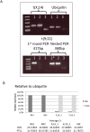
Figure 2. Molecular study of the presence of BCR-ABL1 and t(X;14)-derived fusion transcripts in diagnostic samples corresponding to the two malignancies.
(A) End-point qualitative PCR for the translocation product fusions using primers that map up and down stream of the breakpoints. For t(9;22) also nested PCR was performed. Sample legend: 1 – CML (diagnostic PB sample), 2 – T-NHL (diagnostic PB sample), 3 – H2O, * – 1st round PCR product diluted 1:100 before being used as template in nested PCR. The bands visible in the no-template control lanes are due to primer dimer. (B) Quantitative PCR for the fusion gene transcripts. A graph of relative expression and tables of average Cts are shown. The relative quantity was calculated considering the amount of BCR-ABL1, normalised to ubiquitin, as 100% in the diagnostic CML sample and t(X;14) translocation product 100% in the diagnostic lymphoma sample.
Figure 3
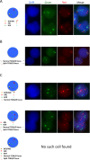
Figure 3. FISH characterisation of diagnostic CML and PTCL samples using the commercial probes that recognise the BCR/ABL fusion or the TCRA/D split.
(A) Ideogram and a typical example of the pattern observed in pathological nuclei hybridised with the Vysis LSI BCR/ABL DC/DF probe showing one fusion, two green and one red signals corresponding to the loss of the reciprocal ABL locus. (B) Ideogram and a typical example of the pattern seen in lymphoma cells the bear the t(X;14) detected as split in the TCRA/D locus. A normal cell with intact 14q locus as testified by the vicinity of the red and green signals and an abnormal cell in which one of the two red/green pairs is separated due to the 14q break are shown. (C) Experiments performed combining the two probes. Big red signal corresponds to the ABL locus, the big green one to the BCR locus, whist the little red and green hybridisation spots correspond to the TCRA/D locus. Ideograms and examples of hybridisation are shown as well as an ideogram of an inexistent cell bearing both translocations.

References

    1. Jabbour E. & Kantarjian H. Chronic myeloid leukemia: 2014 update on diagnosis, monitoring, and management. Am J Hematol 89, 547–556 (2014). - PubMed
    1. Yang K. & Fu L. W. Mechanisms of resistance to BCR-ABL TKIs and the therapeutic strategies: A review. Crit Rev Oncol Hematol 93, 277–92 (2014). - PubMed
    1. Verma D. et al. Malignancies occurring during therapy with tyrosine kinase inhibitors (TKIs) for chronic myeloid leukemia (CML) and other hematologic malignancies. Blood 118, 4353–4358 (2011). - PMC - PubMed
    1. Duman B. B., Paydas S., Disel U., Besen A. & Gurkan E. Secondary malignancy after imatinib therapy: eight cases and review of the literature. Leuk Lymphoma 53, 1706–1708 (2012). - PubMed
    1. Maerki J., Katava G., Siegel D., Silberberg J. & Bhattacharyya P. K. Unusual case of simultaneous presentation of plasma cell myeloma, chronic myelogenous leukemia, and a jak2 positive myeloproliferative disorder. Case Rep Hematol 2014, 738428, 10.1155/2014/738428 (2014). - DOI - PMC - PubMed

Publication types

MeSH terms