Review
. 2015 Nov;40(11):701-714.
doi: 10.1016/j.tibs.2015.08.006. Epub 2015 Oct 1.

Microhomology-Mediated End Joining: A Back-up Survival Mechanism or Dedicated Pathway?

Affiliations
Review

Microhomology-Mediated End Joining: A Back-up Survival Mechanism or Dedicated Pathway?

Agnel Sfeir et al. Trends Biochem Sci. 2015 Nov.

Abstract

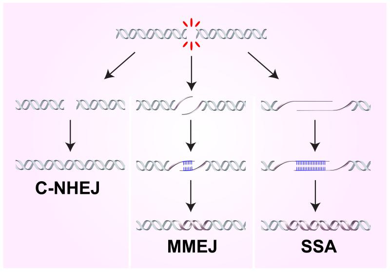
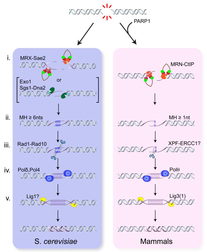
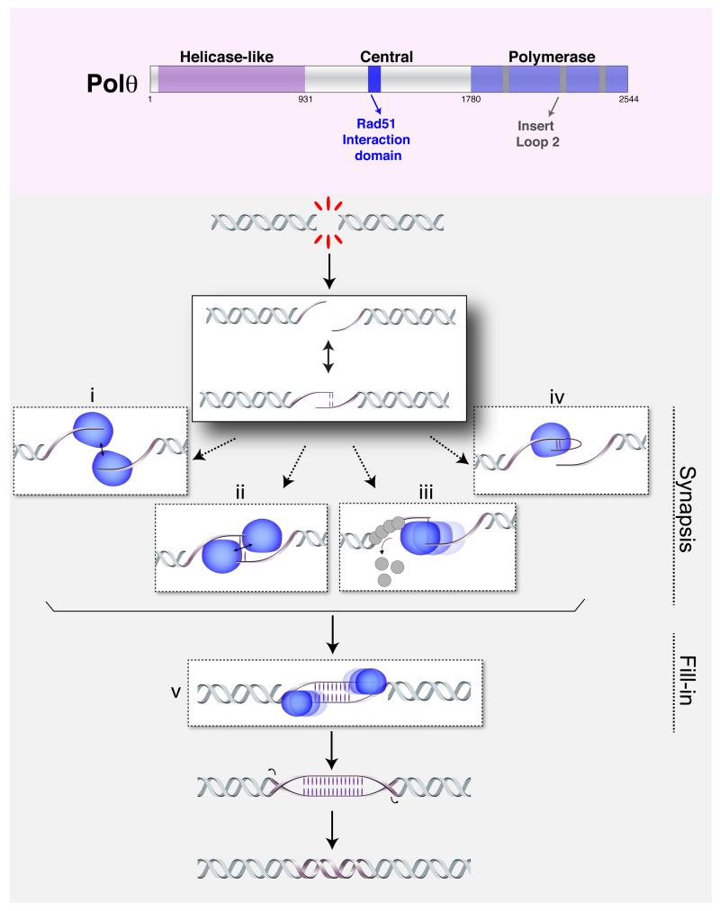
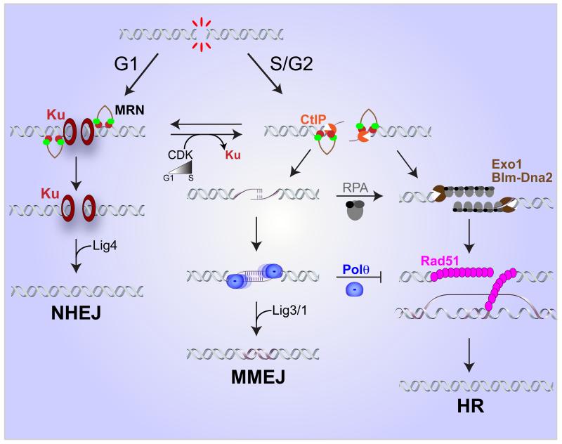
DNA double-strand breaks (DSBs) disrupt the continuity of chromosomes and their repair by error-free mechanisms is essential to preserve genome integrity. Microhomology-mediated end joining (MMEJ) is an error-prone repair mechanism that involves alignment of microhomologous sequences internal to the broken ends before joining, and is associated with deletions and insertions that mark the original break site, as well as chromosome translocations. Whether MMEJ has a physiological role or is simply a back-up repair mechanism is a matter of debate. Here we review recent findings pertaining to the mechanism of MMEJ and discuss its role in normal and cancer cells.

Keywords: DNA Polθ; MMEJ; chromosomal translocations; end joining; homologous recombination; microhomology.


Figures

Figure 1
Figure 1. Mechanisms of end joining
Classical non-homologous end-joining (C-NHEJ) involves no homology or only 1-4 nucleotides of homology at the junction; microhomology mediated end-joining (MMEJ) requires 1-16 nt of homology internal to the ends to align them for repair; and single-stranded annealing (SSA) involves annealing between more extensive homologies provided by direct repeats flanking the DSB. MMEJ and SSA are both highly mutagenic due to loss of one repeat and the intervening sequence.
Figure 2
Figure 2. Mechanistic basis for MMEJ
The illustration highlights the similarities and differences between budding yeast and mammals. The mechanism in both organisms involves i. end resection, ii. annealing of MHs, iii. flap removal, iv. fill-in synthesis, and v. ligation (see text for details). In S. cerevisiae, Exo1 or Sgs1-Dna2 can substitute for MRX and Sae2 to initiate end resection at endonuclease-induced DSBs.
Figure 3
Figure 3. Model for Polθ activity
Upper panel depicts the different domains of Polθ including the helicase-like domain at the N terminus, the polymerase domain at the C terminus, and the central domain that contains the Rad51 interaction motif. The polymerase domain contains an insert loop 2 that is necessary for DNA lesion bypass, ssDNA extension, and MMEJ. In the lower panel, we propose a model for the mechanism by Polθ (in blue) promotes MMEJ Nucleolytic processing of a DSB exposes MH, which could drive annealing of single stranded overhangs. Polθ potentially employ its different domains in order to promote formation and/or stabilization of the annealed intermediate. For example, its dimerization will tether DNA ends together (i) or even stabilize spontaneously annealed ends (ii). The ATPase domain could potentially displace ssDNA-binding proteins (iii). Lastly, the polymerase could extend resected ends, potentially in a snap-back mechanism, thereby generating additional MH (iv). In addition to promoting synapsis, Polθ activity is required for fill-in synthesis (v), a crucial step during MMEJ.
Figure 4
Figure 4. Repair pathway choice
The choice between C-NHEJ, MMEJ and HR is regulated by multiple factors and influenced by the cell cycle. Low CDK activity in G1 enables Ku-dependent NHEJ activity. Increased CDK in S/G2 promotes limited resection by MRN-CtIP and results in a concomitant loss of Ku binding to DSBs. This enables both MMEJ and HR to act. Extensive resection by Exo1 or Blm-Dna2, binding of RPA and subsequent loading of Rad51 are required for HDR. By contrast, the activity of Polθ favors MMEJ, and inhibits HR. In S. cerevisiae RPA inhibits MMEJ by counteracting annealing between MH.

References

    1. Chiruvella KK, et al. Repair of double-strand breaks by end joining. Cold Spring Harbor perspectives in biology. 2013;5:a012757. - PMC - PubMed
    1. Boulton SJ, Jackson SP. Saccharomyces cerevisiae Ku70 potentiates illegitimate DNA double-strand break repair and serves as a barrier to error-prone DNA repair pathways. The EMBO journal. 1996;15:5093–5103. - PMC - PubMed
    1. Ma JL, et al. Yeast Mre11 and Rad1 proteins define a Ku-independent mechanism to repair double-strand breaks lacking overlapping end sequences. Molecular and cellular biology. 2003;23:8820–8828. - PMC - PubMed
    1. Yu X, Gabriel A. Ku-dependent and Ku-independent end-joining pathways lead to chromosomal rearrangements during double-strand break repair in Saccharomyces cerevisiae. Genetics. 2003;163:843–856. - PMC - PubMed
    1. Robert I, et al. Parp1 facilitates alternative NHEJ, whereas Parp2 suppresses IgH/c-myc translocations during immunoglobulin class switch recombination. The Journal of experimental medicine. 2009;206:1047–1056. - PMC - PubMed

Publication types