. 2015 Oct 6:5:14753.
doi: 10.1038/srep14753.

Kinetic characterization of trans-proteolytic activity of Chikungunya virus capsid protease and development of a FRET-based HTS assay

Affiliations

Kinetic characterization of trans-proteolytic activity of Chikungunya virus capsid protease and development of a FRET-based HTS assay

Megha Aggarwal et al. Sci Rep. .

Abstract

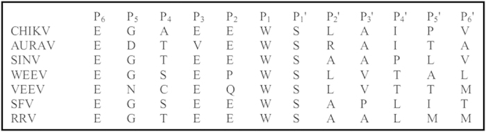
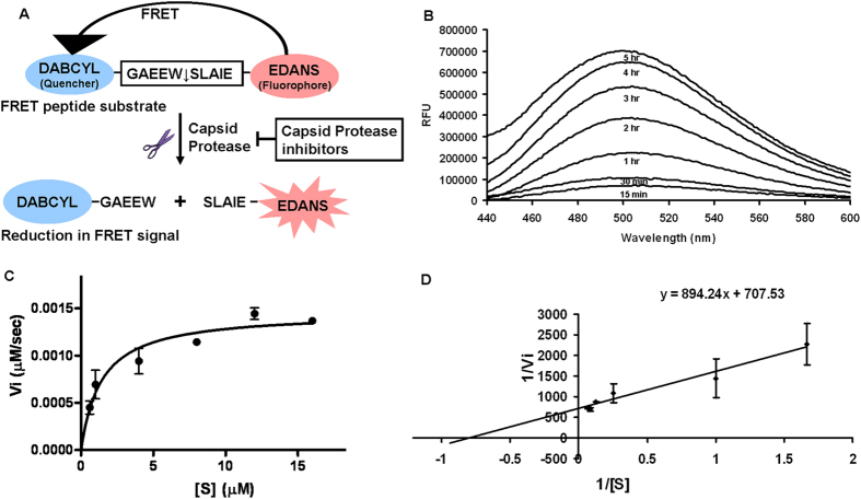
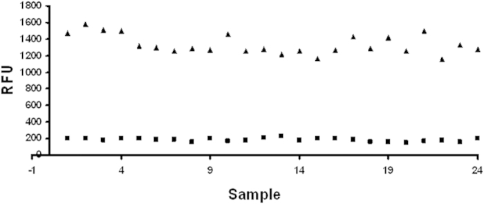
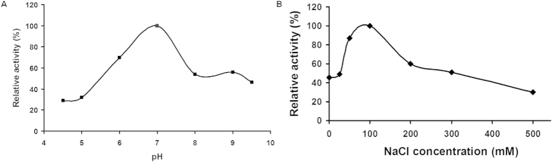
Chikungunya virus (CHIKV) capsid protein (CVCP) is a serine protease that possesses cis-proteolytic activity essential for the structural polyprotein processing and plays a key role in the virus life cycle. CHIKV being an emerging arthropod-borne pathogenic virus, is a public health concern worldwide. No vaccines or specific antiviral treatment is currently available for chikungunya disease. Thus, it is important to develop inhibitors against CHIKV enzymes to block key steps in viral reproduction. In view of this, CVCP was produced recombinantly and purified to homogeneity. A fluorescence resonance energy transfer (FRET)-based proteolytic assay was developed for high throughput screening (HTS). A FRET peptide substrate (DABCYL-GAEEWSLAIE-EDANS) derived from the cleavage site present in the structural polyprotein of CVCP was used. The assay with a Z' factor of 0.64 and coefficient of variation (CV) is 8.68% can be adapted to high throughput format for automated screening of chemical libraries to identify CVCP specific protease inhibitors. Kinetic parameters Km and kcat/Km estimated using FRET assay were 1.26 ± 0.34 μM and 1.11 × 10(3) M(-1) sec(-1) respectively. The availability of active recombinant CVCP and cost effective fluorogenic peptide based in vitro FRET assay may serve as the basis for therapeutics development against CHIKV.


Figures

Figure 1
Figure 1. The peptide sequence of the CP from different alphaviruses showing the residues P6-P6’ surrounding the scissile bond residues Trp and Ser.
Figure 2
Figure 2. SDS-PAGE and gel filtration profile of CVCP.
Both (A) inactive and (B) active CVCP shows the protein purified to homogeneity. The size-exclusion chromatography results suggest the monomeric nature of both the proteins. Lane 1, molecular-weight markers (kDa); lane 2, pellet containing insoluble protein fraction; lane 3, supernatant containing soluble protein fraction; lane 4–6, purified CVCP.
Figure 3
Figure 3. In vitro FRET based assay.
(A) A FRET based protease assay has been developed for the determination of CVCP activity. The DABCYL (Quencher) and EDANS (fluorophore) are attached at the N and C terminus of the peptide respectively. The donor EDANS produces the signal upon excitation that gets quenched by DABCYL and thus shows FRET. In the presence of protease, the cleavage takes place and the fluorophore and quencher gets separated from each other, which results in the reduction of FRET signal. In the presence of protease inhibitors, the FRET should remain unaltered. (B) In vitro trans proteolytic assay was performed in 20 mM HEPES pH 7.0 buffer and the fluorescence was measured at different time points. The fluorescence was measured till 5 hrs and the increase in fluorescence was observed. All the values are the average of triplicate data. The values were normalized using the reaction performed in the similar conditions with no enzyme. (C) To perform the kinetic studies of CVCP, different concentrations of the substrate ranging from 0.6 μM to 16 μM were used and initial velocities were calculated for all substrate concentrations. The kinetic data were fitted into the Michealis-Menten equation. (D) The lineweaver-burk plot was formed and the intercept and slope were calculated according to the equation y = mx + c. From these, the values of Vmax and Km were determined and the kcat was also calculated by dividing Vmax with the enzyme concentration.
Figure 4
Figure 4. Z’ factor analysis.
The scatter plot showing the positive control (triangle shaped) and negative control (square shaped) data for the Z’ factor calculation. 24 samples were used for computing the means and standard deviations. The experiment was performed in triplicate.
Figure 5
Figure 5. The effect of pH and NaCl concentration on the enzymatic activity was observed.
(A) Using the buffers of pH ranging from 4.5 to 9.5, the pH optimum was calculated for the proteolytic activity. The relative activity was calculated at different pHs by taking the activity at pH 7.0 as 100%. (B) The activity at NaCl concentration of 100 mM was taken as 100% and the relative activity for other NaCl concentrations was calculated. All the readings are the average of the triplicate data and the values are normalized by using readings obtained from the reaction containing no enzyme.
Figure 6
Figure 6. Influence of the glycerol on CVCP activity.
(A) The effect of increase in glycerol concentration (0 to 50%) in the reaction buffer on the enzymatic activity of CVCP has been observed and the relative activity was measured by taking the activity at 20% glycerol as 100%. The graph represents the average data obtained from three experiments. (B) CVCP 3D homology model (magenta) with glycerol in the S1 specificity pocket was generated and superimposed on the crystal structure of AVCP with bound P1 residue (green) (PDB ID: 4AGK). The overall structure shows the presence of two subdomains; the active site is present at the interface of these two subdomains (active site residues are shown in sticks). The S1 specificity pocket is present in the vicinity of the active site (shown in circle) to which the P1 residue Trp267 of native AVCP binds. (C) The zoom-in surface view of the S1 pocket shows that glycerol (blue) in the CVCP structure binds incisively at the position where conserved P1 residue Trp (red) binds in AVCP. The active site is shown in blue color.

References

    1. Larrieu S. et al. Factors associated with persistence of arthralgia among Chikungunya virus-infected travellers: report of 42 French cases. J Clin Virol 47, 85–88 (2010). - PubMed
    1. Ross R. W. The Newala epidemic: III. The virus: isolation, pathogenic properties and relationship to the epidemic. J Hyg (Lond) 54, 177–191 (1956). - PMC - PubMed
    1. Ligon B. L. Reemergence of an unusual disease: the chikungunya epidemic. Semin Pediatr Infect Di 17, 99–104 (2006). - PMC - PubMed
    1. Powers A. M. & Logue C. H. Changing patterns of chikungunya virus: re-emergence of a zoonotic arbovirus. J Gen Virol 88, 2363–2377 (2007). - PubMed
    1. Sergon K. et al. Seroprevalence of chikungunya virus (CHIKV) infection on Lamu Island, Kenya, October 2004. Am J Trop Med Hyg 78, 333–337 (2008). - PubMed

Publication types

MeSH terms

LinkOut - more resources