

Identification of functional networks associated with cell death in the retina of OXYS rats during the development of retinopathy
- PMID: 26440064
- PMCID: PMC4825736
- DOI: 10.1080/15384101.2015.1080399
Identification of functional networks associated with cell death in the retina of OXYS rats during the development of retinopathy
Abstract
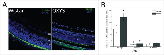
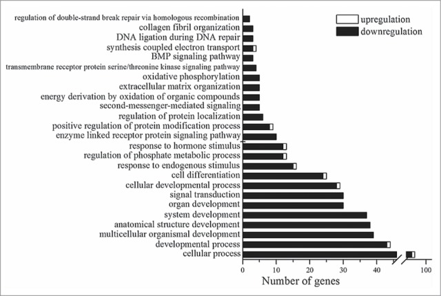
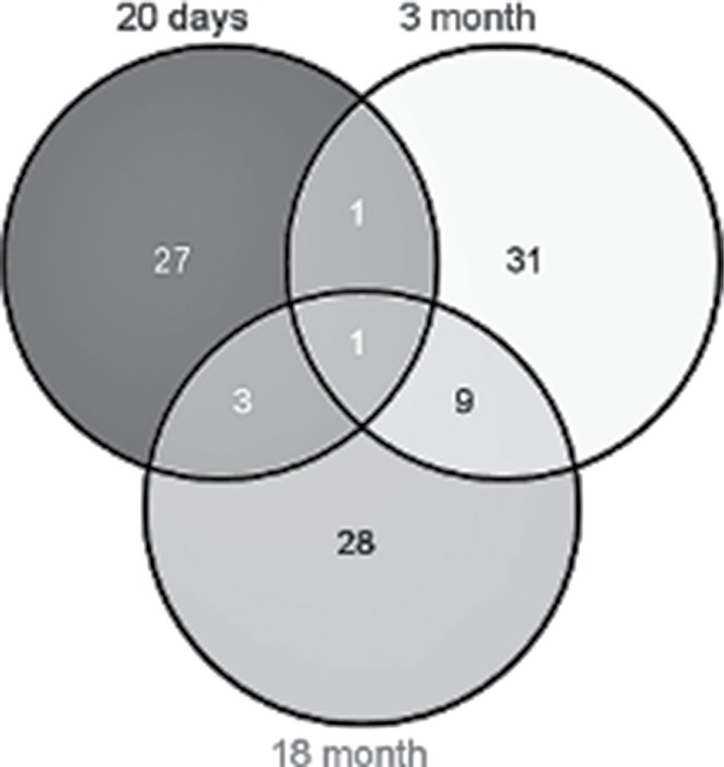
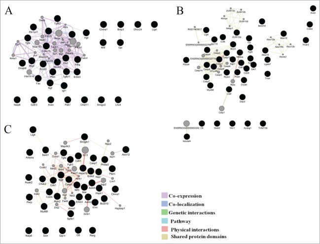
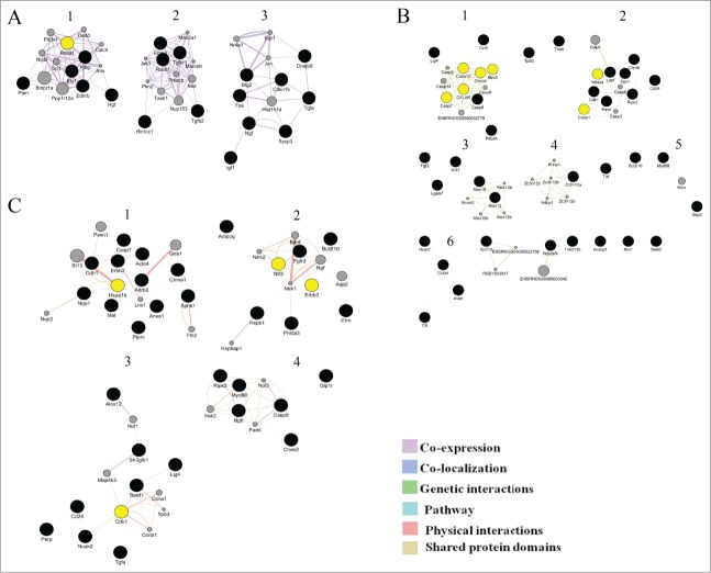
Age-related macular degeneration (AMD) is a major cause of blindness in developed countries, and the molecular pathogenesis of early events in AMD is poorly understood. Senescence-accelerated OXYS rats develop AMD-like retinopathy. The aim of this study was to explore the differences in retinal gene expression between OXYS and Wistar (control) rats at age 20 d and to identify the pathways of retinal cell death involved in the OXYS retinopathy initiation and progression. Retinal mRNA profiles of 20-day-old OXYS and Wistar rats were generated at the sequencing read depth 40 mln, in triplicate, using Illumina GAIIx. A terminal deoxynucleotidyl transferase-mediated deoxyuridine triphosphate nick end labeling (TUNEL) assay was performed to measure the apoptosis level. GeneMANIA was used to construct interaction networks for differentially expressed (DE) apoptosis-related genes at ages 20 d and 3 and 18 months. Functional analysis was suggestive of a developmental process, signal transduction, and cell differentiation as the most enriched biological processes among 245 DE genes at age 20 d An increased level of apoptosis was observed in OXYS rats at age 20 d but not at advanced stages. We identified functional clusters in the constructed interaction networks and possible hub genes (Rasa1, cFLAR, Birc3, Cdk1, Hspa1b, Erbb3, and Ntf3). We also demonstrated the significance of the extrinsic apoptotic pathway at preclinical, early, and advanced stages of retinopathy development. Besides the cell death signaling pathways, immune system-related processes and lipid-metabolic processes showed overrepresentation in the clusters of all networks. These characteristics of the expression profile of the genes functionally associated with apoptosis may contribute to the pathogenesis of AMD-like retinopathy in senescence-accelerated OXYS rats.
Keywords: OXYS rats; RNA-Seq; age-related macular degeneration; aging; apoptosis; cell death; retinal transcriptome.
Figures
Similar articles
-
Immunohistochemical localization of NGF, BDNF, and their receptors in a normal and AMD-like rat retina.BMC Med Genomics. 2019 Mar 13;12(Suppl 2):48. doi: 10.1186/s12920-019-0493-8. BMC Med Genomics. 2019. PMID: 30871541 Free PMC article.
-
Rat retinal transcriptome: effects of aging and AMD-like retinopathy.Cell Cycle. 2013 Jun 1;12(11):1745-61. doi: 10.4161/cc.24825. Epub 2013 May 6. Cell Cycle. 2013. PMID: 23656783 Free PMC article.
-
Application of quantitative trait locus mapping and transcriptomics to studies of the senescence-accelerated phenotype in rats.BMC Genomics. 2014;15 Suppl 12(Suppl 12):S3. doi: 10.1186/1471-2164-15-S12-S3. Epub 2014 Dec 19. BMC Genomics. 2014. PMID: 25563673 Free PMC article.
-
[The senescence-accelerated oxys rats--a genetic model of premature aging and age-dependent degenerative diseases].Adv Gerontol. 2014;27(2):336-40. Adv Gerontol. 2014. PMID: 25306668 Review. Russian.
-
Senescence-accelerated OXYS rats: a model of age-related cognitive decline with relevance to abnormalities in Alzheimer disease.Cell Cycle. 2014;13(6):898-909. doi: 10.4161/cc.28255. Epub 2014 Feb 17. Cell Cycle. 2014. PMID: 24552807 Free PMC article. Review.
Cited by
-
Mechanisms of Neuronal Death in the Cerebral Cortex during Aging and Development of Alzheimer's Disease-Like Pathology in Rats.Int J Mol Sci. 2019 Nov 11;20(22):5632. doi: 10.3390/ijms20225632. Int J Mol Sci. 2019. PMID: 31717998 Free PMC article.
-
Microvascular contributions to age-related macular degeneration (AMD): from mechanisms of choriocapillaris aging to novel interventions.Geroscience. 2019 Dec;41(6):813-845. doi: 10.1007/s11357-019-00138-3. Epub 2019 Dec 4. Geroscience. 2019. PMID: 31797238 Free PMC article. Review.
-
The glutamate/GABA system in the retina of male rats: effects of aging, neurodegeneration, and supplementation with melatonin and antioxidant SkQ1.Biogerontology. 2022 Oct;23(5):571-585. doi: 10.1007/s10522-022-09983-w. Epub 2022 Aug 15. Biogerontology. 2022. PMID: 35969289
-
Immunohistochemical localization of NGF, BDNF, and their receptors in a normal and AMD-like rat retina.BMC Med Genomics. 2019 Mar 13;12(Suppl 2):48. doi: 10.1186/s12920-019-0493-8. BMC Med Genomics. 2019. PMID: 30871541 Free PMC article.
-
Serum RNA Profile Reflects Fluid Status and Atrophic Retinal Changes in Neovascular Age-Related Macular Degeneration.Int J Mol Sci. 2025 May 19;26(10):4852. doi: 10.3390/ijms26104852. Int J Mol Sci. 2025. PMID: 40429992 Free PMC article.
References
-
- Horie-Inoue K, Inoue S. Genomic aspects of age-related macular degeneration. Biochem Biophys Res Commun 2014; 452:263-75; PMID:25111812; http://dx.doi.org/10.1016/j.bbrc.2014.08.013 - DOI - PubMed
-
- Ambati J, Fowler BJ. Mechanisms of age-related macular degeneration. Neuron 2012; 75:26-39; PMID:22794258; http://dx.doi.org/10.1016/j.neuron.2012.06.018 - DOI - PMC - PubMed
-
- de Almagro MC, Vucic D. Necroptosis: Pathway diversity and characteristics. In Semin Cell Dev Biol 2015; 39:56-62; PMID:25683283; http://dx.doi.org/10.1016/j.semcdb.2015.02.002 - DOI - PubMed
-
- Wright AF, Chakarova CF, El-Aziz MMA, Bhattacharya SS. Photoreceptor degeneration: genetic and mechanistic dissection of a complex trait. Nat Rev Genet 2010; 11:273-84; PMID:20212494; http://dx.doi.org/10.1038/nrg2717 - DOI - PubMed
Publication types
MeSH terms
Substances
LinkOut - more resources
Full Text Sources
Other Literature Sources
Medical
Research Materials
Miscellaneous