. 2015 Oct 6;10(10):e0139464.
doi: 10.1371/journal.pone.0139464. eCollection 2015.

Divergent Evolution of the Transcriptional Network Controlled by Snf1-Interacting Protein Sip4 in Budding Yeasts

Affiliations

Divergent Evolution of the Transcriptional Network Controlled by Snf1-Interacting Protein Sip4 in Budding Yeasts

Constance Mehlgarten et al. PLoS One. .

Abstract

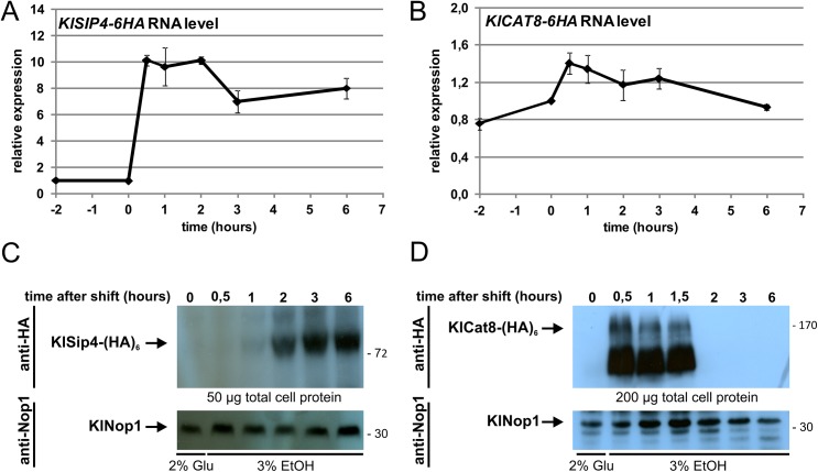
Cellular responses to starvation are of ancient origin since nutrient limitation has always been a common challenge to the stability of living systems. Hence, signaling molecules involved in sensing or transducing information about limiting metabolites are highly conserved, whereas transcription factors and the genes they regulate have diverged. In eukaryotes the AMP-activated protein kinase (AMPK) functions as a central regulator of cellular energy homeostasis. The yeast AMPK ortholog SNF1 controls the transcriptional network that counteracts carbon starvation conditions by regulating a set of transcription factors. Among those Cat8 and Sip4 have overlapping DNA-binding specificity for so-called carbon source responsive elements and induce target genes upon SNF1 activation. To analyze the evolution of the Cat8-Sip4 controlled transcriptional network we have compared the response to carbon limitation of Saccharomyces cerevisiae to that of Kluyveromyces lactis. In high glucose, S. cerevisiae displays tumor cell-like aerobic fermentation and repression of respiration (Crabtree-positive) while K. lactis has a respiratory-fermentative life-style, respiration being regulated by oxygen availability (Crabtree-negative), which is typical for many yeasts and for differentiated higher cells. We demonstrate divergent evolution of the Cat8-Sip4 network and present evidence that a role of Sip4 in controlling anabolic metabolism has been lost in the Saccharomyces lineage. We find that in K. lactis, but not in S. cerevisiae, the Sip4 protein plays an essential role in C2 carbon assimilation including induction of the glyoxylate cycle and the carnitine shuttle genes. Induction of KlSIP4 gene expression by KlCat8 is essential under these growth conditions and a primary function of KlCat8. Both KlCat8 and KlSip4 are involved in the regulation of lactose metabolism in K. lactis. In chromatin-immunoprecipitation experiments we demonstrate binding of both, KlSip4 and KlCat8, to selected CSREs and provide evidence that KlSip4 counteracts KlCat8-mediated transcription activation by competing for binding to some but not all CSREs. The finding that the hierarchical relationship of these transcription factors differs between K. lactis and S. cerevisiae and that the sets of target genes have diverged contributes to explaining the phenotypic differences in metabolic life-style.


Conflict of interest statement

Competing Interests: The authors have declared that no competing interests exist.

Figures

Fig 1
Fig 1. Sequence conservation in the Zn(II)2Cys6 fungal-type DNA-binding domain of Cat8 and Sip4 family members.
(A) Multiple sequence alignment of fungal Zn(II)2Cys6 binuclear cluster motif, linker and coiled-coil regions of Sip4- and Cat8-like proteins derived from various fungal genomic sequences: K. lactis, S. cerevisiae, A. gossypii, C. glabrata, Z. rouxii, L. thermotolerans, V. polyspora, A. nidulans, A. niger and A. oryzae. Identical residues are shown in black boxes and the cysteine residues involved in the coordination of the Zn2+ atoms are marked with an asterisk. The arrow indicates a conserved valine residue following the point of sequence divergence in Cat8 vs. Sip4 as mentioned in the text. (B) Phylogenetic relationship based upon full-length amino acid sequences of Sip4 and Cat8 homologs in selected Ascomycota. The relationship is presented as a phylogram with branch lengths proportional to sequence deviation. Cat8 but no Sip4 homologs are found in Aspergillus spp..
Fig 2
Fig 2. Influence of deletions in KlCAT8 and KlSIP4 on carbon utilization.
K. lactis wild-type (JA6), Klcat8Δ (yIG8), Klsip4Δ (JA6/DS4) and Klcat8ΔKlsip4Δ (yIG8/DS4) strains were pregrown in YP medium with 2% glucose (see Material and Methods) and spotted in serial 10-fold dilutions on minimal plates with 2% of the indicated carbon sources. Plates were incubated at 30°C for 4 days.
Fig 3
Fig 3. Sip4 is involved in expression of carnitine shuttle genes in K. lactis.
(A) Schematic overview of metabolic pathways and key genes essential for the utilization of non-fermentable carbon source ethanol in yeast as found in the yeast genome database [33]. (B) RNA levels of genes related to the carnitine shuttle in sip4Δ, cat8Δ and sip4Δcat8Δ mutants relative to congenic wild-type strains were determined by qRT-PCR in S. cerevisiae (left panel) and K. lactis (right panel). Cultures were shifted from 2% glucose to 3% ethanol medium for 2 hours at an OD600 of 0.8 to 1.0. Gene expression levels were normalized to the reference gene HEM2 and quantified relative to wild-type levels (set to 1.0; dashed line) Data points and error bars represent mean values ± standard deviations obtained with three independent biological samples each measured in technical triplicates. Asterisks indicate statistically significant differences compared to wild-type (t-test; *P < 0.001; ns, not significant). (C) Protein levels of Yat2 in wild-type and mutant strains. In S. cerevisiae (left) the chromosomal YAT2 gene was tagged with a (HA)6-epitope in wild-type and cat8Δ and sip4Δ mutant backgrounds (strains CMY196, CMY198 and CMY197), respectively. In K. lactis the strains JA6 (WT), yIG8 (cat8Δ) and JA6/DS4 (sip4Δ) were transformed with centromeric plasmid pCM68 (KlYAT2-6HA::pKATUC4) or empty vector (pKATUC4). Cultures were grown in glucose or ethanol medium and the indicated amounts of protein extracts were probed by Western blotting with anti-HA antibody (top panel). Nop1, detected with an anti-Nop1 antibody served as loading control (bottom panel). The position of KlYat2-(HA)6 (104.6 kDa) and ScYat2-(HA)6 (110.5 kDa) are indicated by arrows. Numbers refer to molecular markers in kDa.
Fig 4
Fig 4. KlSIP4-6HA and KlCAT8-6HA RNA and protein levels after a shift to ethanol.
(A and B) Time course of KlSIP4-6HA (A) and KlCAT8-6HA (B) gene expression. Wild-type cells expressing KlSip4-(HA)6 (JA6/S4HA) or KlCat8-(HA)6 (JA6/C8HA) were grown in glucose and shifted to ethanol medium at time zero. Samples were taken at the indicated time points, RNA was isolated and qRT-PCR was performed in triplicates. Fold changes relative to the time 0 sample were calculated by the 2−∆∆CT method, normalized to the reference gene KlHEM2. (C and D) Time course of protein levels. In parallel to the RNA preparations (panel A and B) total protein extracts were prepared, separated by SDS-PAGE and analyzed by Western blotting using anti-HA antibody (top panel). Nop1, detected with an anti-Nop1 antibody served as loading control (bottom panel). The position of KlSip4-(HA)6 (89.1 kDa), KlCat8-(HA)6 (166.4 kDa) and KlNop1 (34.8 kDa) are indicated by arrows.
Fig 5
Fig 5. KlSip4-(HA)6 and KlCat8-(HA)6 binding to selected promoter regions.
(A) Schematic overview of potential CSREs (diamonds) matching to the consensus sequences in promoter regions of putative target genes. The fragments amplified with promoter-specific (orange bars) and ORF-specific primer pairs (blue bars) for ChIP-qPCR analysis are indicated. Scale is 200 bp for one graduation. Positions are given relative to the ATG start codon (with +1). Blue diamonds indicate sequences conforming to the loose consensus sequence 5’- CGGNNNNNNGGN-3‘, purple diamonds to the more specific consensus sequence 5’-CGGNTKAAWGGN-3’. (B) CSREs in promoters of KlSip4 target genes and their distance from the ATG translation initiation site. A sequence logo for K. lactis was created using the Weblogo resource (http://weblogo.berkeley.edu/logo.cgi). [35]. The CSRE consensus sequence of S. cerevisiae [34] is shown for comparison. (C and D) ChIP-qPCR results indicating binding of KlSip4-(HA)6 (C) and KlCat8-(HA)6 (D) to selected promoters in strains JA6/S4HA and JA6/C8HA shifted to ethanol for 3 hours or 30 minutes, respectively. ORF-fragments amplified with specific primers pairs served as control for background binding and KlACT1 for normalization to input-DNA. (Reference to mock ChIP with the untagged strain gave similar results as reference to inputs.) Data points and error bars represent mean values ± standard deviations obtained with three independent biological samples each measured in duplicates. The experiment was performed twice with similar results. Asterisks indicate statistically significant differences compared to input-DNA (t-test; *P<0.05; **P<0.01;***P < 0.001; ns, not significant).
Fig 6
Fig 6. Influence of Klsip4 and Klcat8 mutations on KlSIP4 promoter activity.
The KlSIP4 promoter was fused to the β-glucuronidase reporter gene on plasmid pLS2GUS, which was transformed into wild-type cells (JA6) and isogenic mutants Klsip4Δ (JA6/DS4), Klcat8Δ (yIG8) and Klsnf1Δ (JSD1R4), respectively. Transformants were pregrown in 2% glucose medium overnight and then shifted to medium with 3% glycerol, 2% ethanol or without any carbon source for 5 hours. β-glucuronidase activity (mU/mg protein) was assayed in whole cell extracts in three independent measurements and is given relative to the activity in ethanol-grown wild-type cells (100%) determined in parallel in each experiment. Mean values and standard deviation for the three measurements are presented.
Fig 7
Fig 7. The basal control region (BCR) and CSRELAC4 regulate transcription of the LAC4 gene.
(A) Intergenic region between the divergently transcribed genes LAC12 and LAC4 containing the basal control region (BCR, shaded in dark gray) and CSRELAC4 (black box). Four binding sites for the transcription activator KlGal4 are indicated by light grey boxes. Distances are given relative to the LAC4 ATG. (B) Non-induced LAC4 mRNA levels in wild-type and a BCR deletion mutant (ΔBCR; strain JA6/LR2). Total RNA was isolated from cells grown in 3% glycerol and analyzed by quantitative S1 nuclease mapping as described previously [39]. Histone H3 RNA (HHT1, lower lanes) served as loading control.(C) Transcription activation function of CSRELAC4. Non-induced LAC4 expression was determined by β-galactosidase measurements in extracts from wild-type (JA6), JA6/LR2 (ΔBCR) and JA6/LR2K (ΔBCR::CSRELAC4) strains grown in synthetic complete medium containing 2% glucose, 2% acetate or 2% ethanol. (D) Dependence of CSRELAC4–protein complex formation on KlCAT8 and KlSIP4. 10, 20 or 40 μg of S100 protein extracts from wild-type (JA6), sip4Δ (JA6/DS4), cat8Δ (yIG8) or sip4Δcat8Δ (yIG8/DS4) cells grown in synthetic complete medium with 3% glycerol were used for EMSA with a labeled CSRELAC4 oligonucleotide (~80,000 cpm).
Fig 8
Fig 8. Comparison of the SNF1-Cat8-Sip4 regulatory network in K. lactis and S. cerevisiae.
Schematic representation of regulation by the CSRE-binding transcription factors Cat8 and Sip4 for selected orthologous genes. The cartoons symbolize the similarity in the DNA-binding domains (blue ovals), and the differences in the rest of the proteins (blue/yellow rectangles).

Similar articles

Cited by

References

    1. Towler MC, Hardie DG (2007) AMP-activated protein kinase in metabolic control and insulin signaling. Circ Res 100: 328–341. - PubMed
    1. Carling D, Mayer FV, Sanders MJ, Gamblin SJ (2011) AMP-activated protein kinase: nature's energy sensor. Nat Chem Biol 7: 512–518. nchembio.610 [pii]; 10.1038/nchembio.610 - DOI - PubMed
    1. Hardie DG (2011) AMP-activated protein kinase: an energy sensor that regulates all aspects of cell function. Genes Dev 25: 1895–1908. 25/18/1895 [pii]; 10.1101/gad.17420111 - DOI - PMC - PubMed
    1. Gowans GJ, Hardie DG (2014) AMPK: a cellular energy sensor primarily regulated by AMP. Biochem Soc Trans 42: 71–75. BST20130244 [pii]; 10.1042/BST20130244 - DOI - PMC - PubMed
    1. Hardie DG (2011) AMP-activated protein kinase: a cellular energy sensor with a key role in metabolic disorders and in cancer. Biochem Soc Trans 39: 1–13. BST0390001 [pii]; 10.1042/BST0390001 - DOI - PubMed

Publication types

MeSH terms

Substances