. 2015 Nov 5;17(5):585-96.
doi: 10.1016/j.stem.2015.08.019. Epub 2015 Oct 1.

AMPK Protects Leukemia-Initiating Cells in Myeloid Leukemias from Metabolic Stress in the Bone Marrow

Affiliations

AMPK Protects Leukemia-Initiating Cells in Myeloid Leukemias from Metabolic Stress in the Bone Marrow

Yusuke Saito et al. Cell Stem Cell. .

Abstract

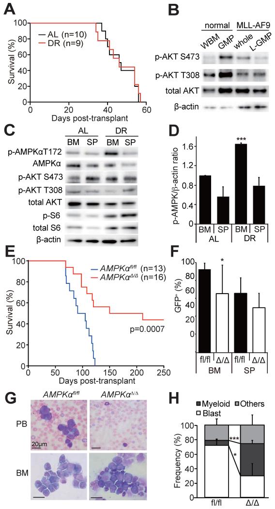
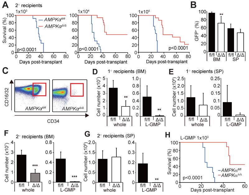
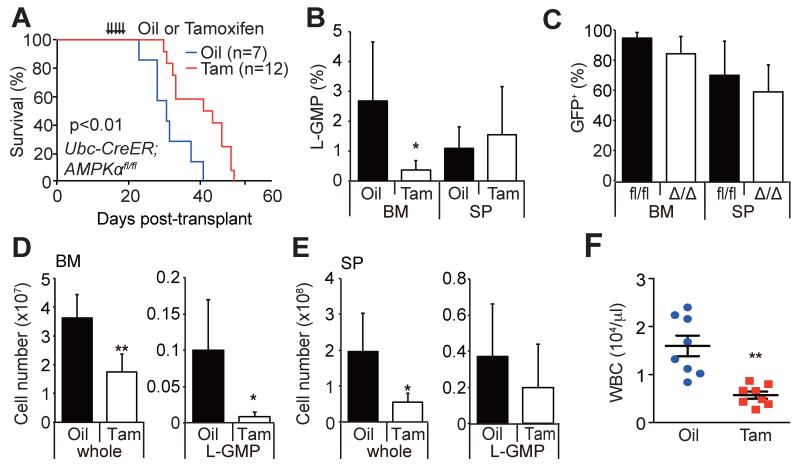
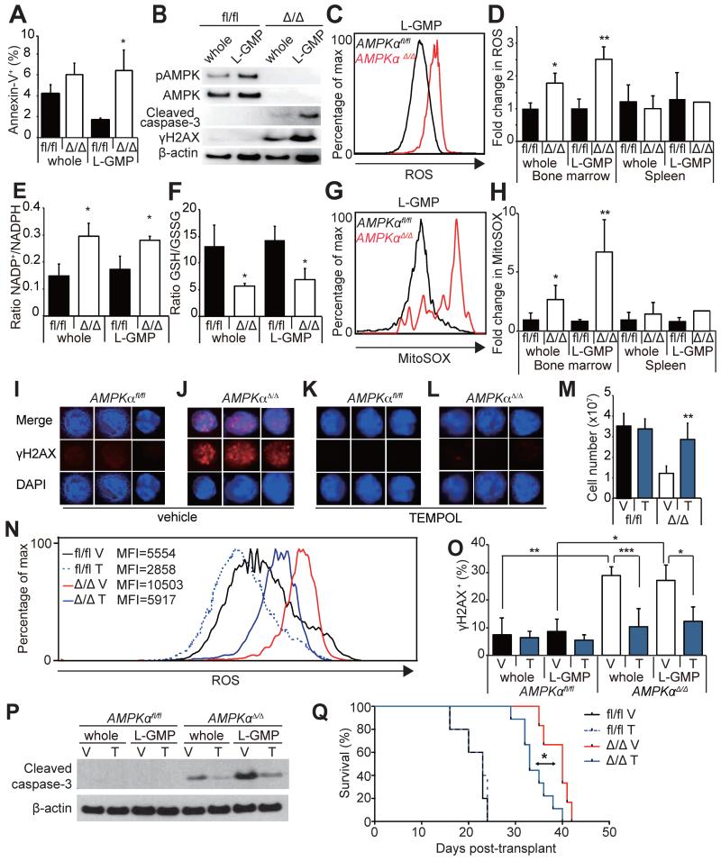
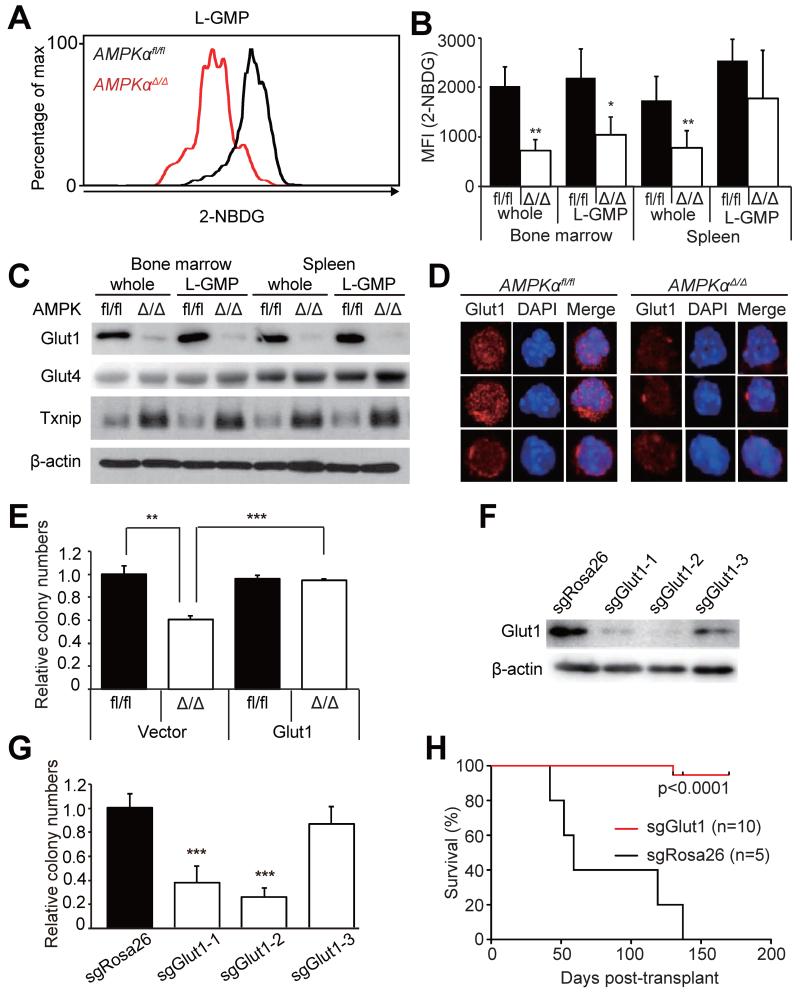
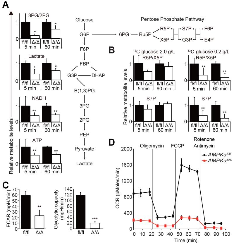
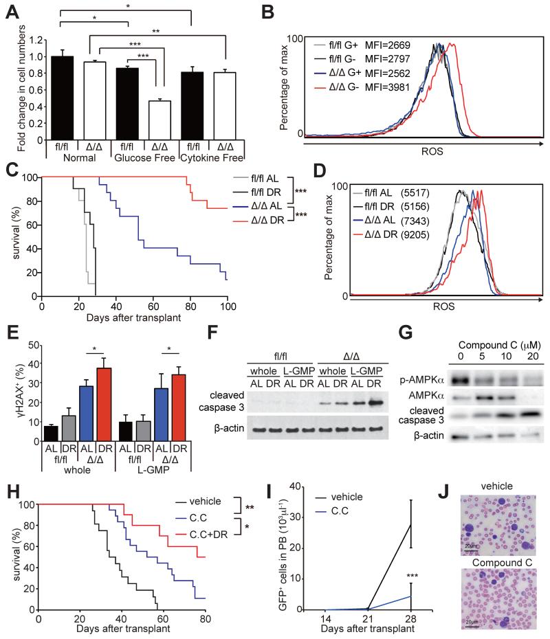
How cancer cells adapt to metabolically adverse conditions in patients and strive to proliferate is a fundamental question in cancer biology. Here we show that AMP-activated protein kinase (AMPK), a metabolic checkpoint kinase, confers metabolic stress resistance to leukemia-initiating cells (LICs) and promotes leukemogenesis. Upon dietary restriction, MLL-AF9-induced murine acute myeloid leukemia (AML) activated AMPK and maintained leukemogenic potential. AMPK deletion significantly delayed leukemogenesis and depleted LICs by reducing the expression of glucose transporter 1 (Glut1), compromising glucose flux, and increasing oxidative stress and DNA damage. LICs were particularly dependent on AMPK to suppress oxidative stress in the hypoglycemic bone marrow environment. Strikingly, AMPK inhibition synergized with physiological metabolic stress caused by dietary restriction and profoundly suppressed leukemogenesis. Our results indicate that AMPK protects LICs from metabolic stress and that combining AMPK inhibition with physiological metabolic stress potently suppresses AML by inducing oxidative stress and DNA damage.

Keywords: AML; AMPK; DNA damage; ROS; dietary restriction; glycolysis; leukemia-initiating cells; metabolic stress.


Figures

Figure 1
Figure 1. AMPK is activated in AML cells upon DR and promotes leukemogenesis
(A) Secondary recipients of 1,000 MLL-AF9-induced AML cells were either fed ad libitum (AL, n=10) or subjected to dietary restriction (DR, n=9). DR did not extend the survival of AML recipient mice. (B) Immunoblotting was performed on freshly isolated whole bone marrow cells (WBM) and GMPs from non-leukemic mice, and whole AML cells (sorted GFP+ cells) and L-GMPs from leukemic mice, all fed AL. PI3K pathway, as determined by phosphorylation of Akt, was not activated in AML cells compared to normal progenitors. (C) Immunoblotting of freshly isolated AML cells from AL or DR mice revealed that AMPK was highly activated in the bone marrow (BM), but not the spleens (SP), of DR mice. (D) Quantification of the results shown in (C). p-AMPK signals were normalized to the signals from β-actin. (E) Survival of mice after transplanting MLL-AF9 transduced AMPKαfl/fl (n=13) or AMPKαΔ/Δ cells (n=16, three independent experiments). (F) Frequencies of GFP+ AML cells in the bone marrow or the spleens of leukemic mice shown in (E). (G) Wright-giemsa staining of peripheral blood (PB) or the bone marrow (BM) samples from AML mice revealed fewer blasts in the recipients of AMPKαΔ/Δ AML. (H) Quantification of the cell types in the bone marrow. In all figures, data represent mean±standard deviation; *, p<0.05; **, p<0.005; and ***, p<0.0005 by Student’s t-test, except for comparison of the survival curves in which the significance was accessed by a log-rank test. See also Figure S1.
Figure 2
Figure 2. AMPK deletion suppresses leukemogenesis and depletes LICs
(A) Secondary transplantation of 103, 104, 105 GFP+ AML cells revealed significantly delayed onset of leukemogenesis by AMPKαΔ/Δ cells. (B) Frequencies of GFP+ AML cells in the bone marrow or the spleens of leukemic mice shown in (A). (C) Representative FACS profiles gated on lineage Sca-1c-kit+cells show reduced CD34+CD16/32+ L-GMPs (red box) after AMPK deletion. (D and E) Total numbers of L-GMPs and whole AML cells in the bone marrow and spleens of primary recipients. (F and G) Secondary recipients of AMPKαΔ/Δ AML cells had significantly reduced total numbers of whole AML cells and L-GMPs in the bone marrow (F), and L-GMPs in the spleens (G). (H) 103 L-GMPs isolated from primary recipients of AMPKαfl/fl and AMPKαΔ/Δ AML were transplanted into secondary recipient mice. AMPKαΔ/Δ L-GMPs had delayed onset of leukemogenesis compared to AMPKαfl/fl L-GMPs, indicating that AMPK deletion impairs L-GMP function. See also Figure S2.
Figure 3
Figure 3. AMPK is required within established leukemias to maintain LICs
(A and B) Conditional deletion of AMPK from established AMLs by treating mice carrying MLL-AF9-transduced Ubc-Cre-ERT2; AMPKαfl/fl AML cells with tamoxifen led to significant extension of survival (A) and reduced frequency of L-GMP in the bone marrow but not in the spleens (B). (C) Acute AMPK deletion did not affect the frequencies of GFP+ AML cells in the bone marrow or in the spleens. (D and E) Acute AMPK deletion significantly reduced the numbers of L-GMPs in the bone marrow but not in the spleens, and reduced the numbers of whole AML cells in these two tissues (n=7). (F) AMPK deletion by tamoxifen treatment reduced WBC in the peripheral blood compared to oil-treated mice (n=8). See also Figure S3.
Figure 4
Figure 4. AMPK deletion induces oxidative stress and DNA damage in LICs
(A) AMPK deletion significantly increased the frequencies of Annexin-V+ L-GMPs in the bone marrow (n=3). (B) Western blotting of AMPKαfl/fl and AMPKαΔ/Δ AML cells and L-GMPs from the bone marrow revealed increased cleaved caspase-3 consistent with the increased cell death, as well as increased γH2AX indicating increased DNA damage. (C and D) AMPK deletion increased the levels of ROS in whole AML cells and L-GMPs in the bone marrow but not in the spleens (n=5). (E and F) Deletion of AMPK from L-GMPs and whole AML cells from the bone marrow increased the NADP+/NADPH ratio and reduced the GSH/GSSG ratio consistent with the increased oxidative stress (n=3). (G and H) AMPK deletion also increased the levels of mitochondrial superoxide (as indicated by the MitoSOX staining) in whole AML cells and L-GMPs in the bone marrow but not in the spleens (n=5). (I-L) Immunofluorescence staining of freshly isolated bone marrow L-GMPs with anti-γH2AX antibodies revealed that AMPK deletion (J) increased DNA damage, which was significantly suppressed by treating the mice with an antioxidant TEMPOL (L). Scale bars indicate 5 μm. Quantification of I-L is shown in O (V: vehicle, T: TEMPOL, n=5). (M) TEMPOL treatment increased the numbers of AMPKαΔ/Δ AML cells in the bone marrow, and reduced ROS levels in bone marrow L-GMPs (N). MFI indicates the mean fluorescence intensity of each sample. (P) Western blotting of AMPKαfl/fl and AMPKαΔ/Δ AML cells and L-GMPs isolated from the bone marrow of mice subjected to TEMPOL or vehicle treatment revealed that the levels of cleaved caspase-3 in AMPKαΔ/Δ AML cells and L-GMPs were partially reduced by TEMPOL treatment. (Q) 104 GFP+ AML cells from secondary recipients were transplanted and recipients treated with either control vehicle or TEMPOL. TEMPOL treatment slightly but significantly accelerated the onset of leukemogenesis by AMPKαΔ/Δ AML cells.
Figure 5
Figure 5. AMPK regulates glucose uptake through Glut1
(A) A representative histogram showing reduced 2-NBDG uptake by AMPKαΔ/Δ bone marrow L-GMPs in vivo. (B) AMPK deletion significantly reduced glucose uptake of whole AML cells in the bone marrow and spleens, and L-GMPs in the bone marrow but not in the spleens (n=5). Immunoblotting (C) and immunostaining (D) assays revealed that AMPK deletion reduced the protein levels of Glut1, but not Glut4, in whole AML cells and L-GMPs, concomitant with increased Txnip1 levels. Scale bars indicate 5 μm. (E) Overexpression of Glut1 substantially rescued the defective clonogenicity of AMPKαΔ/Δ AML cells (n=4). (F) Immunoblotting against Glut1 using gene-edited AML cells revealed that Glut1 sgRNA clones 1 and 2 reduced Glut1 protein levels, whereas clone 3 had little effects. (G) Clonogenecity of the gene-edited cells as in (F). Glut1 sgRNA clones 1 and 2 significantly reduced clonogenicity, whereas clone 3 did not (n=4). (H) AML cells expressing sgRosa26 (n=5) or sgGlut1 (clone 1, n=5; clone 2, n=5) were transplanted. Deletion of Glut1 by sgRNAs significantly attenuated leukemogenesis. See also Figure S4.
Figure 6
Figure 6. AMPK regulates glucose metabolism
(A) 13C-glucose flux analysis performed on freshly isolated AMPKαfl/fl and AMPKαΔ/Δ AML cells exposed to 2.0 g/L 13C-glucose for the indicated time revealed significantly reduced 13C incorporation into glycolysis intermediates 3PG/2PG and lactate, in addition to NADH and ATP. The amounts of 13C-labeled metabolites were normalized to the values of AMPKαfl/fl cells (n=3). Schematic of glycolysis and the pentose phosphate pathway (PPP) is shown on right. (B) 13C-glucose flux analysis was performed as in (A) with either 2.0 g/L (left panels) or 0.2 g/L glucose (right panels) to access the glucose flux through PPP (n=3). (C) Basal ECAR and ECAR after oligomycin treatment were significantly reduced in freshly isolated AMPKαΔ/Δ AML cells compared to AMPKαfl/fl AML cells (n=3). (D) Extracellular Flux analysis revealed that AMPKαfl/fl AML cells have significantly reduced OCR (n=3). See also Figure S5.
Figure 7
Figure 7. AMPK inhibition sensitized AML to dietary restriction
(A) In vitro glucose deprivation, but not cytokine deprivation, significantly reduced survival of AMPKαΔ/Δ AML cells during a 24-hour culture (n=4). (B) Representative histograms showing increased ROS levels in AMPKαΔ/Δ AML cells after glucose deprivation in vitro. G+ and G− indicates cultured with or without glucose, respectively. (C) Secondary recipients of 104 AMPKαfl/fl (n=10 per group) and AMPKαΔ/Δ AML cells (n=15 per group) were either fed AL or subjected to DR and their survival monitored. DR did not extend the survival of AMPKαfl/fl AML recipients, but did significantly extend the survival of AMPKαΔ/Δ AML recipients. (D) Representative histograms showing that DR further increased the higher ROS levels of AMPKαΔ/Δ L-GMPs (n=3). (E) The frequencies of whole AML cells and L-GMPs positive for γH2AX examined by immunofluorescence assay. (F) Immunoblotting assay using AMPKαfl/fl and AMPKαΔ/Δ whole AML cells and L-GMPs isolated from the bone marrow of the recipient mice subjected to AL or DR. (G) Immunoblotting assay of primary AML cells treated with compound C. (H) Secondary recipient mice of 103 MLL-AF9 AML cells were fed AL or subjected to DR after transplantation. 7 days after transplantation, mice were treated with 4 mg/kg of compound C (C.C) or control vehicle. Compound C treatment significantly extended the survival, and this was further extended when combined with DR (vehicle; n=16, C.C; n=18, C.C+DR; n=10). (I and J) Compound C treatment significantly reduced the total numbers of GFP+ AML cells (I) and the frequencies of blasts (J) in peripheral blood (vehicle; n=5, C.C; n=8). Scale bars indicate 20 μm. See also Figure S6.

Comment in

Similar articles

Cited by

References

    1. Almeida A, Moncada S, Bolanos JP. Nitric oxide switches on glycolysis through the AMP protein kinase and 6-phosphofructo-2-kinase pathway. Nat Cell Biol. 2004;6:45–51. - PubMed
    1. Barnes K, Ingram JC, Porras OH, Barros LF, Hudson ER, Fryer LG, Foufelle F, Carling D, Hardie DG, Baldwin SA. Activation of GLUT1 by metabolic and osmotic stress: potential involvement of AMP-activated protein kinase (AMPK) J Cell Sci. 2002;115:2433–2442. - PubMed
    1. Birsoy K, Possemato R, Lorbeer FK, Bayraktar EC, Thiru P, Yucel B, Wang T, Chen WW, Clish CB, Sabatini DM. Metabolic determinants of cancer cell sensitivity to glucose limitation and biguanides. Nature. 2014;508:108–112. - PMC - PubMed
    1. Bochner BR, Siri M, Huang RH, Noble S, Lei XH, Clemons PA, Wagner BK. Assay of the multiple energy-producing pathways of mammalian cells. PLoS ONE. 2011;6:e18147. - PMC - PubMed
    1. Bonnet D, Dick JE. Human acute myeloid leukemia is organized as a hierarchy that originates from a primitive hematopoietic cell. Nat Med. 1997;3:730–737. - PubMed

Publication types

MeSH terms

Substances

Associated data