. 2015 Oct;20(10):106001.
doi: 10.1117/1.JBO.20.10.106001.

Multispectral tissue characterization for intestinal anastomosis optimization

Affiliations

Multispectral tissue characterization for intestinal anastomosis optimization

Jaepyeong Cha et al. J Biomed Opt. 2015 Oct.

Abstract

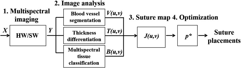
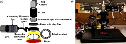
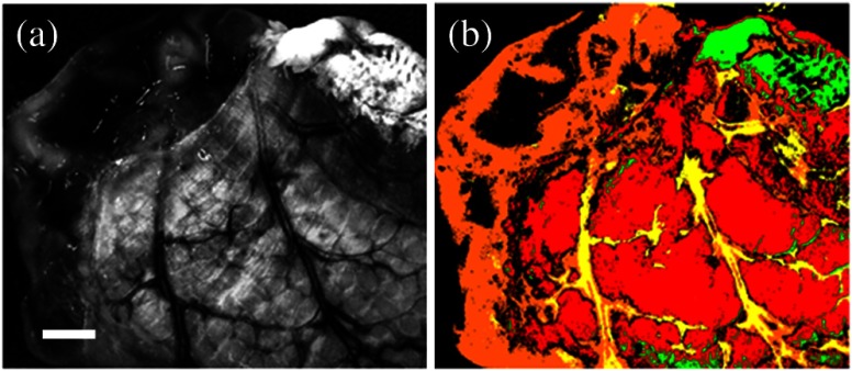
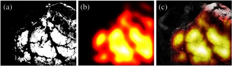
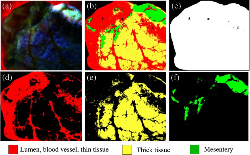
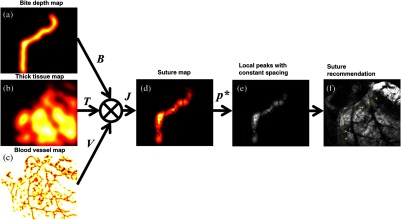
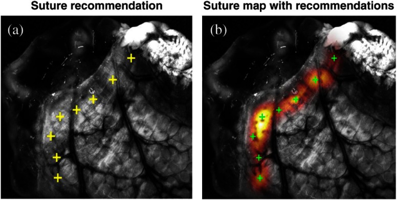
Intestinal anastomosis is a surgical procedure that restores bowel continuity after surgical resection to treat intestinal malignancy, inflammation, or obstruction. Despite the routine nature of intestinal anastomosis procedures, the rate of complications is high. Standard visual inspection cannot distinguish the tissue subsurface and small changes in spectral characteristics of the tissue, so existing tissue anastomosis techniques that rely on human vision to guide suturing could lead to problems such as bleeding and leakage from suturing sites. We present a proof-of-concept study using a portable multispectral imaging (MSI) platform for tissue characterization and preoperative surgical planning in intestinal anastomosis. The platform is composed of a fiber ring light-guided MSI system coupled with polarizers and image analysis software. The system is tested on ex vivo porcine intestine tissue, and we demonstrate the feasibility of identifying optimal regions for suture placement.


Figures

Fig. 1
Fig. 1
System block diagram to create recommendations for optimal suture placements.
Fig. 2
Fig. 2
(a) Schematic of the multispectral imaging (MSI) system and (b) photo of system implementation.
Fig. 3
Fig. 3
Different spectral band images of a tissue sample with (a)–(c) and without cross-polarization, (d)–(f) demonstrating surface reflection removal. Arrows indicate features of blood vessels in red color (a) and (d) and the revealed subsurface features in yellow color. (c) and (f) White scale bars: 2 mm.
Fig. 4
Fig. 4
(a) Blood vessel segmentation using Frangi 2-D filter. (b) Blood vessel segmentation result (red) image overlay on the single-band image at 470 nm. (c) Blood vessel map V(u,v) created by Gaussian filter smoothing of the output of the Frangi 2-D filter. (d) Image overlay of inverted vessel map (inverted for better visualization) on the single-band reflectance image of the intestine.
Fig. 5
Fig. 5
(a) Digital photograph of three bovine colon tissues with different specified thicknesses (units in mm). (b) Thickness differentiation using the SAM method, with a thickness-corresponding colormap. The mesentery is indicated as a different tissue in green color. Black arrows: thicker tissue areas; white scale bar: 10 mm; and colormap unit: mm.
Fig. 6
Fig. 6
(a) Representative single-band reflectance image at 470 nm. (b) Thickness differentiation using the SAM method, with a thickness-corresponding colormap. The red color shows the thicker layer, and the orange color shows the thinner layer. Tissue classification of the mesentery (green) and blood vessels (yellow) was performed prior to the thickness analysis. White scale bar: 2 mm.
Fig. 7
Fig. 7
(a) Thickness binary map as evaluated by the SAM algorithm. (b) Smoothed thickness map T(u,v). Larger values (brighter) denote areas with thicker tissue, which are better suited for suture placement. (c) Overlay of thickness map over a single-band image for better visualization.
Fig. 8
Fig. 8
Multispectral tissue classification: (a) composite image created from three spectral band images; (b) classified image using the supervised classification algorithm; (c) background color-matching image (black); (d) image showing the vulnerable tissue (lumen, blood vessels, and thin tissue regions) (red); (e) image showing the thick tissue regions (yellow); and (f) image showing the mesentery (green).
Fig. 9
Fig. 9
Multispectral image analysis facilitates segmentation of (a) outer layer of the lumen (serosa) and (b) inner layers of the lumen (mucosa and submucosa layers). (c) The cut edge is automatically extracted by pixelwise multiplication of a dilated map of outer and inner layers of the lumen. (d) A bite depth map B(u,v) is generated by translating and smoothing the cut edge by 1.5 times the thickness of the tissue. (e) Overlay of the bite depth map on a single-band image for better visualization.
Fig. 10
Fig. 10
Suture map and suture placement recommendations: (a) bite-depth map B(u,v); (b) thick tissue map T(u,v); (c) blood vessel map V(u,v); (d) combined map J(u,v); (e) selection of local peaks with equal-space consistency constraint; and (f) an overlay image of the recommended suture locations.
Fig. 11
Fig. 11
(a) Magnified view of the suture placement recommendations and (b) colormap overlay of the suture map provided to the surgeon to overrule recommendations and choose other acceptable regions.

References

    1. Wesier T. G., et al. , “An estimation of the global volume of surgery: a modeling strategy based on available data,” Lancet 372(9633), 139–144 (2008).LANCAO10.1016/S0140-6736(08)60878-8 - DOI - PubMed
    1. Slieker J. C., et al. , “Systematic review of the technique of colorectal anastomosis,” JAMA Surg. 148(2), 190–201 (2013).10.1001/2013.jamasurg.33 - DOI - PubMed
    1. Marano L., et al. , “Sutureless jejuno-jejunal anastomosis in gastric cancer patients: a comparison with handsewn procedure in a single institute,” BMC Surg. 12(Suppl 1), S27 (2012).10.1186/1471-2482-12-S1-S27 - DOI - PMC - PubMed
    1. Vignali A., et al. , “Factors associated with the occurrence of leaks in stapled rectal anastomoses: a review of 1, 014 patients,” J. Am. Coll. Surg. 185(2), 105–113 (1997).JACSEX10.1016/S1072-7515(97)00018-5 - DOI - PubMed
    1. Neutzling C. B., et al. , “Stapled versus handsewn methods for colorectal anastomosis surgery,” Cochrane Database Syst. Rev. 2, CD003144 (2012).10.1002/14651858.CD003144.pub2 - DOI - PMC - PubMed

Publication types