. 2015 Oct 13;13(2):425-39.
doi: 10.1016/j.celrep.2015.09.004. Epub 2015 Oct 1.

Circulating Th1-Cell-type Tfh Cells that Exhibit Impaired B Cell Help Are Preferentially Activated during Acute Malaria in Children

Affiliations

Circulating Th1-Cell-type Tfh Cells that Exhibit Impaired B Cell Help Are Preferentially Activated during Acute Malaria in Children

Nyamekye Obeng-Adjei et al. Cell Rep. .

Abstract

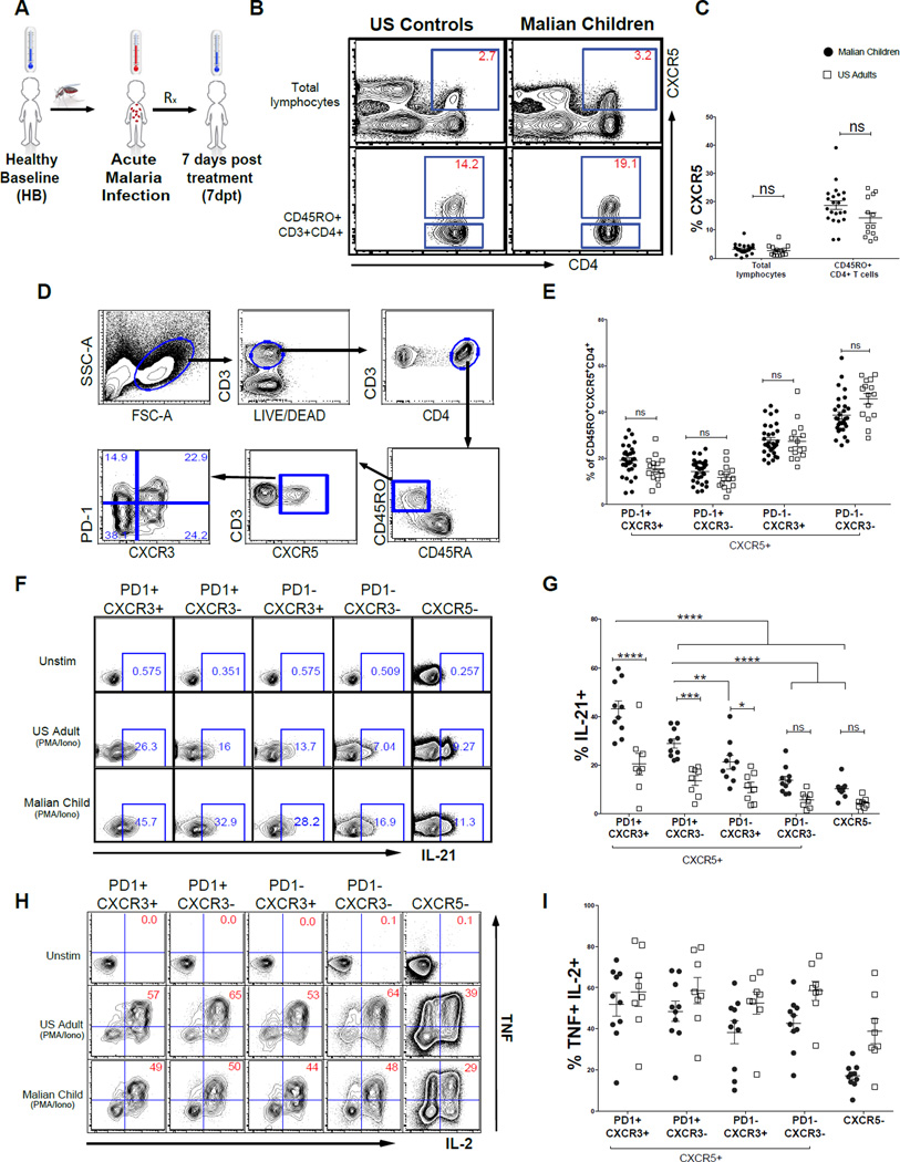
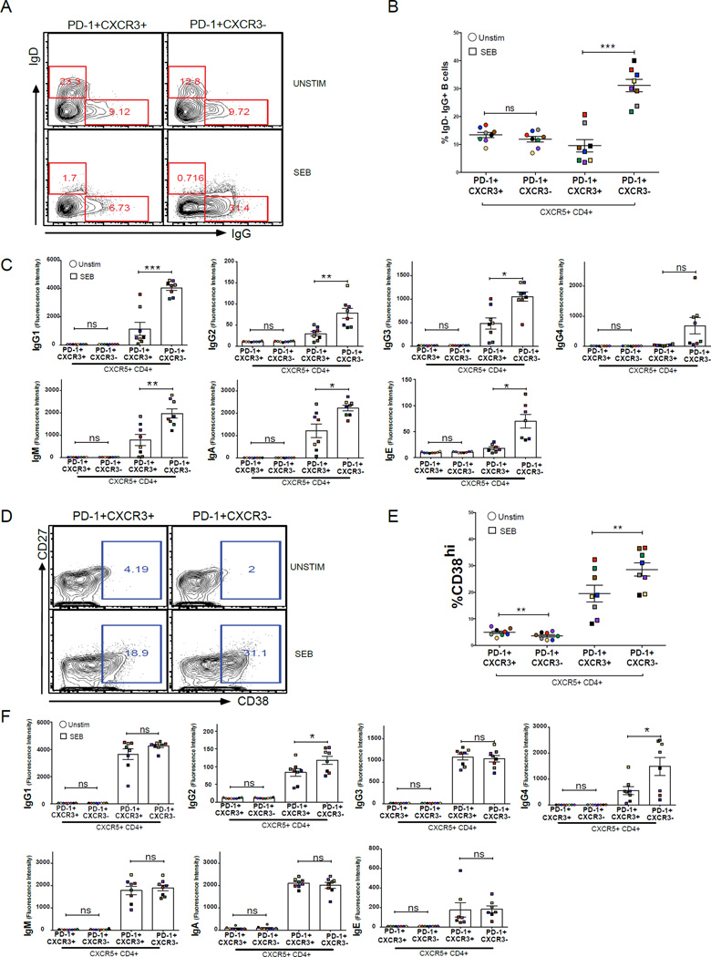
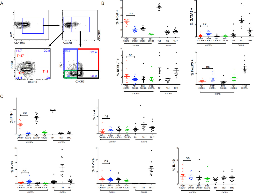
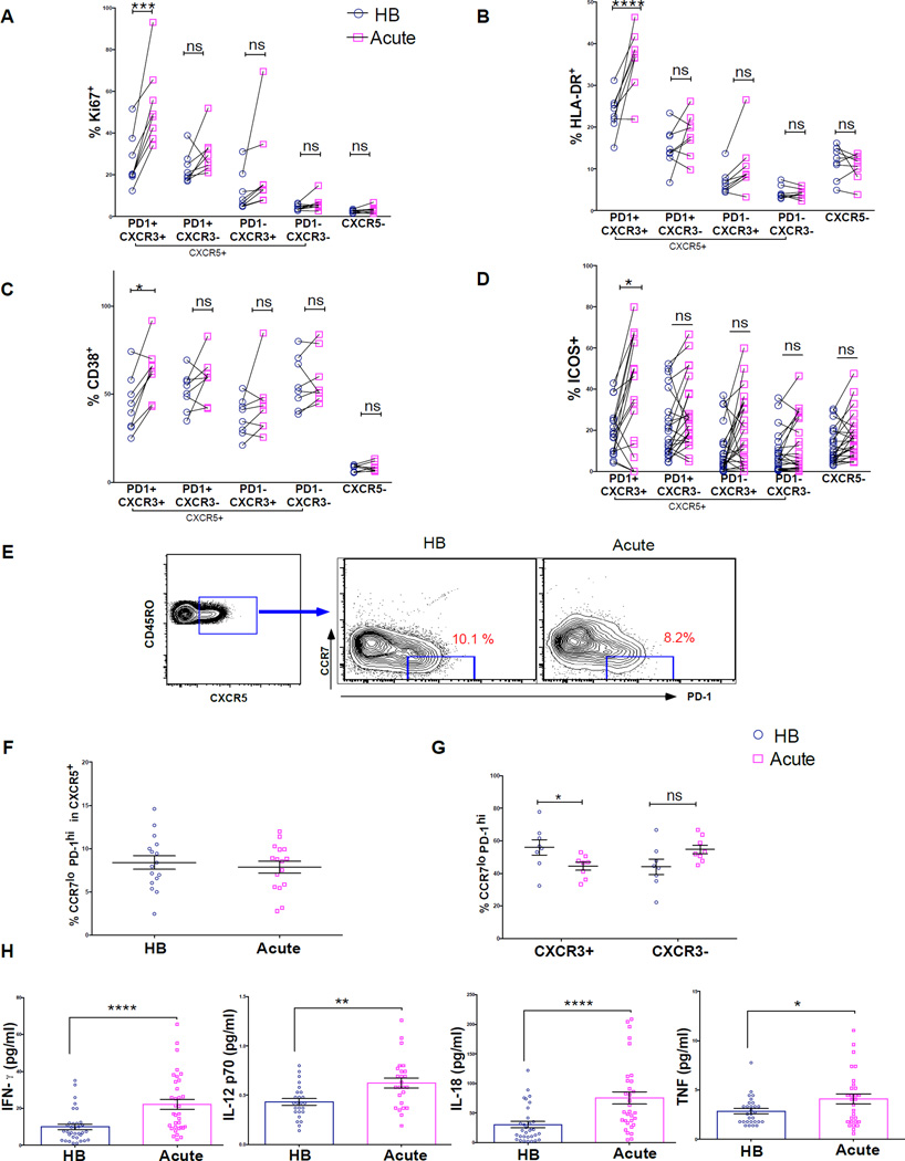
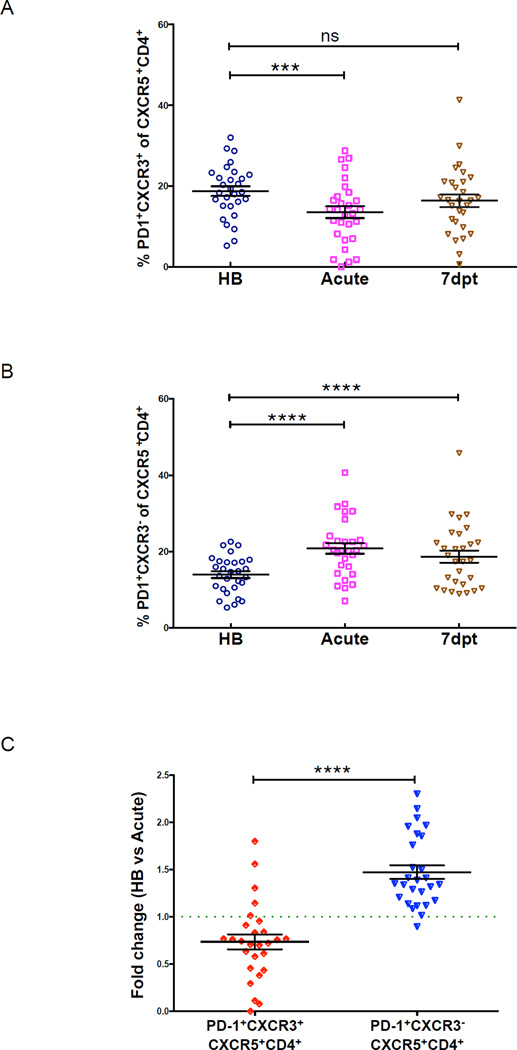
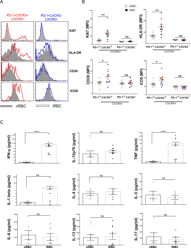
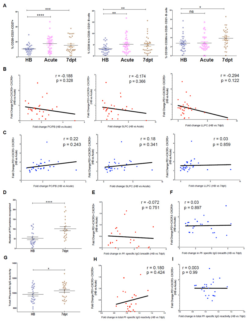
Malaria-specific antibody responses are short lived in children, leaving them susceptible to repeated bouts of febrile malaria. The cellular and molecular mechanisms underlying this apparent immune deficiency are poorly understood. Recently, T follicular helper (Tfh) cells have been shown to play a critical role in generating long-lived antibody responses. We show that Malian children have resting PD-1(+)CXCR5(+)CD4(+) Tfh cells in circulation that resemble germinal center Tfh cells phenotypically and functionally. Within this population, PD-1(+)CXCR5(+)CXCR3(-) Tfh cells are superior to Th1-polarized PD-1(+)CXCR5(+)CXCR3(+) Tfh cells in helping B cells. Longitudinally, we observed that malaria drives Th1 cytokine responses, and accordingly, the less-functional Th1-polarized Tfh subset was preferentially activated and its activation did not correlate with antibody responses. These data provide insights into the Tfh cell biology underlying suboptimal antibody responses to malaria in children and suggest that vaccine strategies that promote CXCR3(-) Tfh cell responses may improve malaria vaccine efficacy.


Conflict of interest statement

The terms of this arrangement have been reviewed and approved by the University of California in accordance with its conflict of interest policies. No other author declares a conflict of interest.

Figures

Figure 1
Figure 1. PD-1 expressing CXCR5+CD4+ T cells from Malian children have features of GC Tfh cells
(A) Longitudinal study design in which blood was collected at the healthy uninfected baseline before the malaria season (HB), during the first acute malaria episode of the ensuing malaria season (acute) and 7 days post-malaria treatment (7dpt). (B and C) The percentage of CXCR5+ cells within total circulating lymphocytes and antigen-experienced CD4+ T cells in Malian children and U.S. adults. (D) Gating strategy for CD45RO+CD45RACXCR5+ CD4+ T helper cell subsets. (E) Distribution of memory CXCR5+ T cell subsets in Malian children and U.S. adults. (F) Intracellular IL-21 production across CXCR5+ subsets with and without PMA/ionomycin in representative Malian child and U.S. adult. (G) Percentage of IL-21 producing cells across CXCR5+ subsets following PMA/ionomycin stimulation in Malian children (n=10) and U.S. adults (n=8). (H) Intracellular IL-2 and TNF production across CXCR5+ subsets with and without PMA/ionomycin in representative Malian child and U.S. adult. (I) Percentage CXCR5+ subsets producing intracellular IL-2 and TNF following PMA/ionomycin stimulation in Malian children (n=10) and U.S. adults (n=10). P values determined by Student’s t test with Bonferroni corrections for multiple comparisons or by ANOVA with Sidak corrections for multiple comparisons where appropriate. ****P<0.0001, ***P<0.001, **P<0.01, *P<0.05, ns=not significant.
Figure 2
Figure 2. PD-1+CXCR3CXCR5+CD4+ Tfh cells are superior to PD-1+CXCR3+CXCR5+CD4+ Tfh cells in providing B cell help
(A–C) PD-1+CXCR3 CXCR5+CD4+ and PD-1+CXCR3+CXCR5+CD4+ Tfh cells from Malian children (n=8) were FACS-sorted and cultured with autologous naïve B cells (CD19+CD21+CD27) with and without SEB. After 12 days, surface expression of IgD and IgG on B cells and secreted Ig levels were measured. (A) Representative plots of IgD and IgG staining on B cells after 12-day co-culture. (B) Percentage of IgDIgG+ B cells after 12-day co-culture. (C) Total IgG1-4, IgM, IgA and IgE levels in supernatants after 12-day co-culture. (D–F) The same FACS-sorted PD-1+CXCR3CXCR5+CD4+ and PD-1+CXCR3+CXCR5+CD4+ Tfh subsets were cultured for 6 days with autologous MBCs (CD19+CD21+CD27+) with and without SEB. After 6 days, surface expression of CD27 and CD38 on B cells and secreted Ig levels were measured. (D) Representative plot of CD27 and CD38 staining on B cells after 6-day co-culture. (E) Percentage of B cells that were CD38hi plasmablasts after 6-day co-culture. (F) Total IgG1-4, IgM, IgA and IgE levels in supernatants after 6-day co-culture. Samples from the same subject are matched by color. P values were determined by paired Student’s t test with Bonferroni adjustments. ****P<0.0001, ***P<0.001, **P<0.01, *P<0.05, ns=not significant.
Figure 3
Figure 3. PD-1+CXCR3+CXCR5+CD4+ Tfh cells are Th1-polarized
(A) Gating strategy for the analysis of CXCR5+ and CXCR5 memory CD4+ T cell subsets. (B) Ex vivo expression of T-bet, GATA-3, RORγT and Foxp3 by CXCR5+ and CXCR5 CD4+ T cell subsets. (C) Intracellular production of indicated cytokines by CXCR5+ and CXCR5 CD4+ T cell subsets following PMA/ionomycin stimulation. Only PD1+CXCR3+ and PD1+CXCR3 were compared and p-values were determined by paired Student’s t test. ****P<0.0001, ***P<0.001, **P<0.01, *P<0.05, ns=not significant.
Figure 4
Figure 4. Acute malaria induces Th1 cytokines and activates the less functional Th1-polarized CXCR3+ Tfh subset
(A–D) Percentage of CXCR5+ subsets expressing the proliferation/activation markers Ki67, CD38, HLA-DR and the co-stimulatory molecule ICOS during acute febrile malaria (acute) compared to the healthy uninfected baseline (HB). (E) Representative FACS plot showing PD-1 and CCR7 expression within antigen-experienced CXCR5+CD4+ T cells in a Malian child at the HB and during acute malaria (F) Percentage CCR7loPD-1hi cells within CXCR5+CD4+ T cells at the HB and during acute malaria. (G) Percentage CCR7loPD-1hi cells within the CXCR3+ and CXCR3 Tfh subsets at the HB and during acute malaria. (H) Plasma cytokine levels at HB and acute. P values determined by paired-design ANOVA with Sidak corrections for multiple comparisons (A-D) or paired Student’s t test with Bonferroni corrections for multiple comparisons where appropriate (F–H). ****P<0.0001, ***P<0.001, **P<0.01, *P<0.05, ns=not significant
Figure 5
Figure 5. PD-1+CXCR3+CXCR5+CD4+ T cells transiently decrease in circulation during acute malaria
Percentage of circulating (A) PD-1+CXCR3+CXCR5+CD4+ and (B) PD-1+CXCR3CXCR5+CD4+ T cells in Malian children at the healthy baseline (HB), during acute malaria (acute) and 7 days post-treatment (7dpt). (C) Fold change in percentage of circulating PD-1+CXCR3+CXCR5+CD4+ and PD-1+CXCR3CXCR5+CD4+ T cells from HB to acute. P values determined by paired-design ANOVA with Tukey adjustments for multiple comparisons where appropriate. ****P<0.0001, ***P<0.001, **P<0.01, *P<0.05, ns=not significant.
Figure 6
Figure 6. P. falciparum-infected RBCs drive a Th1 cytokine response and preferentially activate CXCR3+ Tfh cells
PBMCs collected before the malaria season from healthy uninfected children with prior malaria exposure were stimulated in vitro with P. falciparum-infected red blood cells (iRBCs) or naïve red blood cells (nRBCs) for 5 days. (A) Representative plots showing the expression of Ki67, HLA-DR, CD38 and ICOS in the PD-1+CXCR3+ and PD-1+CXCR3 subsets following stimulation. (B) MFI of Ki67, HLA-DR, CD38 and ICOS in the PD-1+CXCR3+ and PD-1+CXCR3 subsets following stimulation. (C) Cytokine levels in supernatants of stimulated PBMCs. P values determined by paired-design ANOVA with Sidak corrections for multiple comparisons or paired Student’s t test with Bonferroni corrections for multiple comparisons. ****P<0.0001, ***P<0.001, **P<0.01, *P<0.05, ns=not significant.
Figure 7
Figure 7. Tfh cell responses to malaria do not correlate with plasma cell and antibody responses to the same infection
(A) Circulating total PCs (CD20CD21 CD27+) (left), SLPCs (CD20CD21CD27+CD38hi) (middle) and LLPCs (CD20CD21CD27+CD38hiCD138) (right) as a percentage of total CD19+ cells in children at healthy baseline (HB), during acute malaria (acute) and 7 days post-treatment (7dpt). (B) Fold change in CXCR3+ Tfh cells (HB to acute) versus fold change in total PCs (left) and SLPCs (middle) (HB to acute) and LLPCs (right) (HB to 7dpt). (C) Fold change in CXCR3 Tfh cells (HB to acute) versus fold change in total PCs (left) and SLPCs (middle) (HB to acute) and LLPCs (right) (HB to 7dpt). (D) Antibody breadth of IgG response to 1087 P. falciparum antigens at HB and at 7dpt. Fold change in (E) CXCR3+ or (F) CXCR3 Tfh cells from HB to acute versus fold change in breadth of IgG response from HB to 7dpt. (G) Total IgG reactivity to 1087 P. falciparum antigens during HB and 7dpt. Fold change in (H) CXCR3+ or (I) CXCR3 Tfh cells from HB to acute versus fold change in total IgG reactivity to 1087 P. falciparum antigens from HB to 7dpt. Student’s t test and Pearson correlation were used. ****P<0.0001, ***P<0.001, **P<0.01, *P<0.05, ns=not significant.

References

    1. Akiba H, Takeda K, Kojima Y, Usui Y, Harada N, Yamazaki T, Ma J, Tezuka K, Yagita H, Okumura K. The role of ICOS in the CXCR5+ follicular B helper T cell maintenance in vivo. Journal of immunology. 2005;175:2340–2348. - PubMed
    1. Alonso PL, Sacarlal J, Aponte JJ, Leach A, Macete E, Aide P, Sigauque B, Milman J, Mandomando I, Bassat Q, et al. Duration of protection with RTS,S/AS02A malaria vaccine in prevention of Plasmodium falciparum disease in Mozambican children: single-blind extended follow-up of a randomised controlled trial. Lancet. 2005;366:2012–2018. - PubMed
    1. Amanna IJ, Carlson NE, Slifka MK. Duration of humoral immunity to common viral and vaccine antigens. N Engl J Med. 2007;357:1903–1915. - PubMed
    1. Bentebibel SE, Lopez S, Obermoser G, Schmitt N, Mueller C, Harrod C, Flano E, Mejias A, Albrecht RA, Blankenship D, et al. Induction of ICOS+CXCR3+CXCR5+ TH cells correlates with antibody responses to influenza vaccination. Sci Transl Med. 2013;5:176ra132. - PMC - PubMed
    1. Breitfeld D, Ohl L, Kremmer E, Ellwart J, Sallusto F, Lipp M, Forster R. Follicular B helper T cells express CXC chemokine receptor 5, localize to B cell follicles, and support immunoglobulin production. J Exp Med. 2000;192:1545–1552. - PMC - PubMed

Publication types

Supplementary concepts