

Assessing blood granulocyte colony-stimulating factor as a potential biomarker of acute traumatic brain injury in mice and humans
- PMID: 26441136
- PMCID: PMC5873950
- DOI: 10.1016/j.bbi.2015.10.002
Assessing blood granulocyte colony-stimulating factor as a potential biomarker of acute traumatic brain injury in mice and humans
Abstract
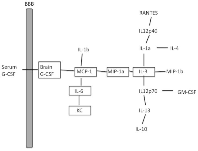
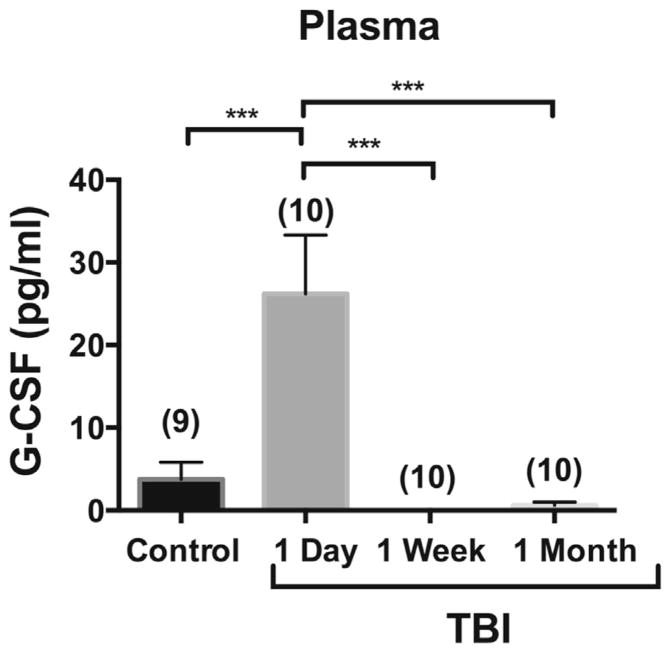
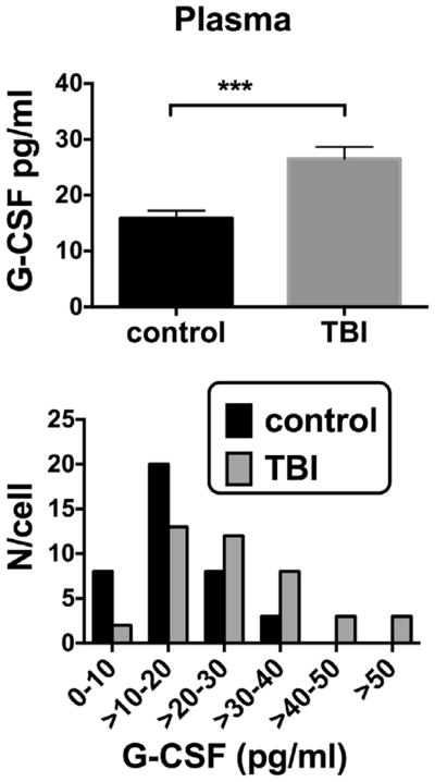
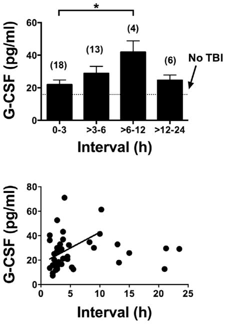
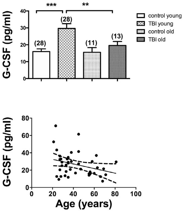
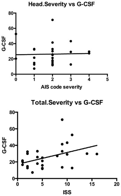
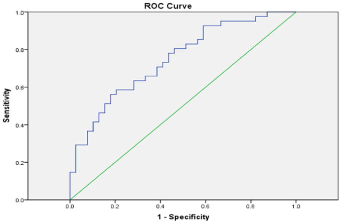
Previous work has found that serum G-CSF was acutely elevated in mice 24h but not one week after controlled cortical impact (CCI). The purpose of this study was to investigate whether blood G-CSF correlates with the elevated brain cytokines in mice after CCI and also if it correlates with traumatic brain injury (TBI) in humans. Here, we found in mice undergoing CCI, a procedure that induces direct injury to the brain, that serum G-CSF correlated directly or indirectly with several brain cytokines, indicating it is a useful marker for the neuroinflammation of TBI. A pilot study in humans (phase I, n=19) confirmed that plasma G-CSF is acutely elevated on day 1 (p<0.001) of TBI and has returned to baseline by one week. In a second human sample (phase II) (n=80), we found plasma G-CSF peaks about 12h after arriving in the emergency department (41.6+/-5.4 pg/ml). Aging was weakly associated (p<0.05) with a less robust elevation in serum G-CSF, but there was no difference with gender. ISS, a measure of total severity of injury, correlated with the degree of elevation in serum G-CSF (r=.419; p<0.05), but severity of head injury (via AIS) did not. The latter may have been because of the statistically narrow range of head injuries among our cases and the high number of cases diagnosed with closed head injury (a non-codable diagnosis). In conclusion, plasma G-CSF may be a useful biomarker of TBI, correlating with neuroinflammation in the animal model and in the human studies with time since injury and total severity of injury. As such, it may be useful in determining whether TBI has occurred within the last 24h.
Keywords: Biomarker; Blood–brain barrier; G-CSF; Neuroinflammation; Traumatic brain injury.
Published by Elsevier Inc.
Figures
References
-
- Acosta SA, Tajiri N, Shinozuka K, Ishikawa H, Sanberg PR, Sanchez-Ramos J, Song S, Kaneko Y, Borlongan CV. Combination therapy of human umbilical cord blood cells and granulocyte colony stimulating factor reduces histopathological and motor impairments in an experimental model of chronic traumatic brain injury. PLoS One. 2014;9:e90953. - PMC - PubMed
-
- Baker SP, O’Neill B. The injury severity score: an update. J Trauma. 1976;16:882–885. - PubMed
-
- Baker SP, O’Neill B, Haddon W, Jr, Long WB. The injury severity score: a method for describing patients with multiple injuries and evaluating emergency care. J Trauma. 1974;14:187–196. - PubMed
-
- Baron RM, Kenny DA. The moderator-mediator variable distinction in social psychological research: conceptual, strategic, and statistical considerations. Pers Soc Psychol. 1986;51:1173–1182. - PubMed
Publication types
MeSH terms
Substances
Grants and funding
LinkOut - more resources
Full Text Sources
Other Literature Sources
Medical
