. 2016 Dec;31(6):472-485.
doi: 10.1111/omi.12140. Epub 2015 Nov 16.

Structure of RagB, a major immunodominant outer-membrane surface receptor antigen of Porphyromonas gingivalis

Affiliations

Structure of RagB, a major immunodominant outer-membrane surface receptor antigen of Porphyromonas gingivalis

T Goulas et al. Mol Oral Microbiol. 2016 Dec.

Abstract

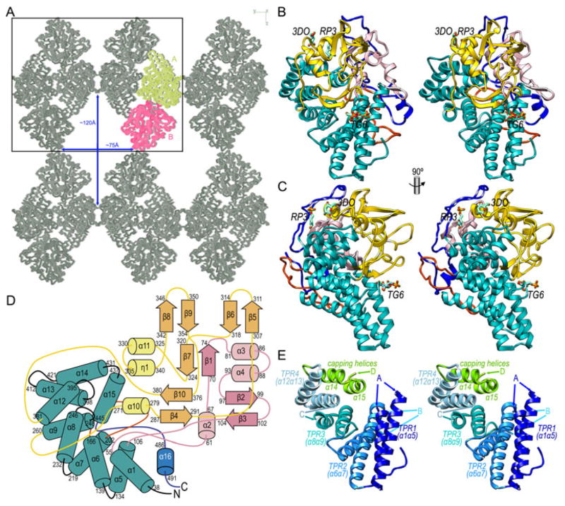
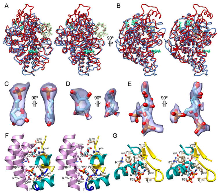
Porphyromonas gingivalis is the main causative agent of periodontitis. It deregulates the inflammatory and innate host immune responses through virulence factors, which include the immunodominant outer-membrane surface receptor antigens A (PgRagA) and B (PgRagB), co-transcribed from the rag pathogenicity island. The former is predicted to be a Ton-dependent porin-type translocator but the targets of this translocation and the molecular function of PgRagB are unknown. Phenomenologically, PgRagB has been linked with epithelial cell invasion and virulence according to murine models. It also acts as a Toll-like receptor agonist and promotes multiple mediators of inflammation. Hence, PgRagB is a candidate for the development of a periodontitis vaccine, which would be facilitated by the knowledge of its atomic structure. Here, we crystallized and solved the structure of 54-kDa PgRagB, which revealed a single domain centered on a curved helical scaffold. It consists of four tetratrico peptide repeats (TPR1-4), each arranged as two helices connected by a linker, plus two extra downstream capping helices. The concave surface bears four large intertwined irregular inserts (A-D), which contribute to an overall compact moiety. Overall, PgRagB shows substantial structural similarity with Bacteroides thetaiotaomicron SusD and Tannerella forsythia NanU, which are, respectively, engaged in binding and uptake of malto-oligosaccharide/starch and sialic acid. This suggests a similar sugar-binding function for PgRagB for uptake by the cognate PgRagA translocator, and, consistently, three potential monosaccharide-binding sites were tentatively assigned on the molecular surface.

Keywords: SusD-like proteins; X-ray crystal structure; periodontitis; sugar-binding proteins; tetratricorepeat proteins.


Figures

Figure 1
Figure 1. Molecular structure of P. gingivalis RagB
(A) Crystal packing of PgRagB in Cα traces viewed along cell axis a. The two molecules in the asymmetric unit, A and B, are shown in yellow and magenta, respectively, symmetry equivalents are in grey. The box delineates the bc plane of the unit cell. Large solvent channels—of maximal dimensions ~75Å and ~120Å traverse the crystal along cell axis a. (B) Ribbon-type plot of PgRagB in cross-eye stereo, showing the TPRs in turquoise, insert A in light pink, insert B in red, insert C in gold, and the C-terminal insert D in dark blue. The three tentative sugar moieties—labeled RP,3 3DO and TG6—are depicted as stick models with carbons in turquoise. (C) Orthogonal view of (B). (D) Topology scheme of PgRagB depicting helices as rods (labeled α1-α16 and η1) and strands as arrows (β1-β10), the numbers of the delimiting residues are shown in each case. Color code similar to (B). (E) Cartoon in stereo depicting only the four tetratrico repeats (TPR1-4) of PgRagB and the capping helices (α14+α15), each shown in one color and labeled. The points of insertion of inserts A, B, C, and D are shown by arrows. Orientation as in (B).
Figure 2
Figure 2. Structural similarities and possible ligands
(A) Superposition in cross-eye stereo of PgRagB (blue ribbon, tentative sugars in turquoise) and BtSusD in its complex with maltoheptaose (red ribbon, sugar moieties in pink; PDB 3CK9; (Koropatkin et al., 2008)). Orientation as in Fig. 1B. (B) Orthogonal view of (A). (C) (2mFobsDFcalc)-type Fourier omit map contoured at 1σ displayed as a purple semi-transparent surface in two orthogonal views around the final refined stick model of tentative ligand RP3 (carbons in light blue). (D) Same as (C) for tentative ligand 3DO. (E) Same as (C) for tentative ligand TG6, modeled as an open keto form with double conformation. (F) Close-up view in stereo of the binding sites of tentative ligands RP3 and 3DO, shown as stick models with carbons in light blue. Molecule A is shown in the colors of Fig. 1B, molecule B in light purple. Side chains participating in ligand binding are shown for their side chains (molecule A, carbons in tan; molecule B, carbons in light purple). (G) Same as (F) for the tentative TG6-binding site.

References

    1. Afonine PV, Grosse-Kunstleve RW, Echols N, et al. Towards automated crystallographic structure refinement with phenix.refine. Acta Crystallogr sect D. 2012;68:352–367. - PMC - PubMed
    1. Anderson KL, Salyers AA. Genetic evidence that outer membrane binding of starch is required for starch utilization by Bacteroides thetaiotaomicron. J Bacteriol. 1989a;171:3199–3204. - PMC - PubMed
    1. Anderson KL, Salyers AA. Biochemical evidence that starch breakdown by Bacteroides thetaiotaomicron involves outer membrane starch-binding sites and periplasmic starch-degrading enzymes. J Bacteriol. 1989b;171:3192–3198. - PMC - PubMed
    1. Bagaitkar J, Williams LR, Renaud DE, et al. Tobacco-induced alterations to Porphyromonas gingivalis-host interactions. Environ Microbiol. 2009;11:1242–1253. - PMC - PubMed
    1. Bakolitsa C, Xu Q, Rife CL, et al. Structure of BT_3984, a member of the SusD/RagB family of nutrient-binding molecules. Acta Crystallogr sect F. 2010;66:1274–1280. - PMC - PubMed

MeSH terms

LinkOut - more resources