Endothelial Mineralocorticoid Receptor Deletion Prevents Diet-Induced Cardiac Diastolic Dysfunction in Females
- PMID: 26441470
- PMCID: PMC4644106
- DOI: 10.1161/HYPERTENSIONAHA.115.06015
Endothelial Mineralocorticoid Receptor Deletion Prevents Diet-Induced Cardiac Diastolic Dysfunction in Females
Abstract
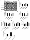
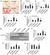
Overnutrition and insulin resistance are especially prominent risk factors for the development of cardiac diastolic dysfunction in females. We recently reported that consumption of a Western diet (WD) containing excess fat (46%), sucrose (17.5%), and high fructose corn syrup (17.5%) for 16 weeks resulted in cardiac diastolic dysfunction and aortic stiffening in young female mice and that these abnormalities were prevented by mineralocorticoid receptor blockade. Herein, we extend those studies by testing whether WD-induced diastolic dysfunction and factors contributing to diastolic impairment, such as cardiac fibrosis, hypertrophy, inflammation, and impaired insulin signaling, are modulated by excess endothelial cell mineralocorticoid receptor signaling. Four-week-old female endothelial cell mineralocorticoid receptor knockout and wild-type mice were fed mouse chow or WD for 4 months. WD feeding resulted in prolonged relaxation time, impaired diastolic septal wall motion, and increased left ventricular filling pressure indicative of diastolic dysfunction. This occurred in concert with myocardial interstitial fibrosis and cardiomyocyte hypertrophy that were associated with enhanced profibrotic (transforming growth factor β1/Smad) and progrowth (S6 kinase-1) signaling, as well as myocardial oxidative stress and a proinflammatory immune response. WD also induced cardiomyocyte stiffening, assessed ex vivo using atomic force microscopy. Conversely, endothelial cell mineralocorticoid receptor deficiency prevented WD-induced diastolic dysfunction, profibrotic, and progrowth signaling, in conjunction with reductions in macrophage proinflammatory polarization and improvements in insulin metabolic signaling. Therefore, our findings indicate that increased endothelial cell mineralocorticoid receptor signaling associated with consumption of a WD plays a key role in the activation of cardiac profibrotic, inflammatory, and growth pathways that lead to diastolic dysfunction in female mice.
Keywords: cardiovascular diseases; diet, Western; insulin resistance; obesity; receptors, mineralocorticoid.
© 2015 American Heart Association, Inc.
Figures


Similar articles
-
Mineralocorticoid receptor blockade prevents Western diet-induced diastolic dysfunction in female mice.Am J Physiol Heart Circ Physiol. 2015 May 1;308(9):H1126-35. doi: 10.1152/ajpheart.00898.2014. Epub 2015 Mar 6. Am J Physiol Heart Circ Physiol. 2015. PMID: 25747754 Free PMC article.
-
Endothelial Mineralocorticoid Receptor Mediates Diet-Induced Aortic Stiffness in Females.Circ Res. 2016 Mar 18;118(6):935-943. doi: 10.1161/CIRCRESAHA.115.308269. Epub 2016 Feb 15. Circ Res. 2016. PMID: 26879229 Free PMC article.
-
Enhanced endothelium epithelial sodium channel signaling prompts left ventricular diastolic dysfunction in obese female mice.Metabolism. 2018 Jan;78:69-79. doi: 10.1016/j.metabol.2017.08.008. Epub 2017 Sep 8. Metabolism. 2018. PMID: 28920862
-
Role of mineralocorticoid receptor activation in cardiac diastolic dysfunction.Biochim Biophys Acta Mol Basis Dis. 2017 Aug;1863(8):2012-2018. doi: 10.1016/j.bbadis.2016.10.025. Epub 2016 Oct 29. Biochim Biophys Acta Mol Basis Dis. 2017. PMID: 27989961 Free PMC article. Review.
-
Role of the vascular endothelial sodium channel activation in the genesis of pathologically increased cardiovascular stiffness.Cardiovasc Res. 2022 Jan 7;118(1):130-140. doi: 10.1093/cvr/cvaa326. Cardiovasc Res. 2022. PMID: 33188592 Free PMC article. Review.
Cited by
-
Myocardial Ischemia and Diabetes Mellitus: Role of Oxidative Stress in the Connection between Cardiac Metabolism and Coronary Blood Flow.J Diabetes Res. 2019 Apr 4;2019:9489826. doi: 10.1155/2019/9489826. eCollection 2019. J Diabetes Res. 2019. PMID: 31089475 Free PMC article. Review.
-
Effects of Bergamot Polyphenols on Mitochondrial Dysfunction and Sarcoplasmic Reticulum Stress in Diabetic Cardiomyopathy.Nutrients. 2021 Jul 20;13(7):2476. doi: 10.3390/nu13072476. Nutrients. 2021. PMID: 34371986 Free PMC article. Review.
-
Pathophysiological Link and Treatment Implication of Heart Failure and Preserved Ejection Fraction in Patients with Chronic Kidney Disease.Biomedicines. 2024 Apr 30;12(5):981. doi: 10.3390/biomedicines12050981. Biomedicines. 2024. PMID: 38790943 Free PMC article. Review.
-
Cellular mechanisms underlying obesity-induced arterial stiffness.Am J Physiol Regul Integr Comp Physiol. 2018 Mar 1;314(3):R387-R398. doi: 10.1152/ajpregu.00235.2016. Epub 2017 Nov 22. Am J Physiol Regul Integr Comp Physiol. 2018. PMID: 29167167 Free PMC article. Review.
-
Mineralocorticoid Receptor and Endothelial Dysfunction in Hypertension.Curr Hypertens Rep. 2019 Sep 4;21(10):78. doi: 10.1007/s11906-019-0981-4. Curr Hypertens Rep. 2019. PMID: 31485760 Free PMC article. Review.
References
-
- Mosca L, Manson JE, Sutherland SE, Langer RD, Manolio T, Barrett-Connor E, Writing Group Cardiovascular disease in women: a statement for healthcare professionals from the American Heart Association. Circulation. 1997;96:2468–2482. - PubMed
-
- Rutter MK, Parise H, Benjamin EJ, Levy D, Larson MG, Meigs JB, Nesto RW, Wilson PW, Vasan RS. Impact of glucose intolerance and insulin resistance on cardiac structure and function: sex-related differences in the Framingham Heart Study. Circulation. 2003;107:448–454. - PubMed
-
- Mihailidou AS, Ashton AW. Cardiac effects of aldosterone: does gender matter? Steroids. 2014;91:32–37. - PubMed
Publication types
MeSH terms
Substances
Grants and funding
LinkOut - more resources
Full Text Sources
Medical

