Review
. 2015 Jul;28(3):125-32.
doi: 10.1293/tox.2015-0025. Epub 2015 Jun 1.

Practical approaches for evaluating adrenal toxicity in nonclinical safety assessment

Affiliations
Review

Practical approaches for evaluating adrenal toxicity in nonclinical safety assessment

Akira Inomata et al. J Toxicol Pathol. 2015 Jul.

Erratum in

  • Errata (Printer's correction).
    [No authors listed] [No authors listed] J Toxicol Pathol. 2016 Jan;29(1):74. Epub 2016 Feb 17. J Toxicol Pathol. 2016. PMID: 26989306 Free PMC article.

Abstract

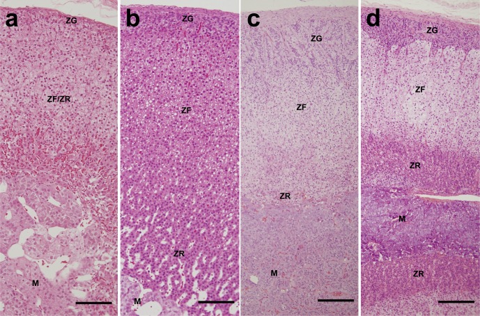
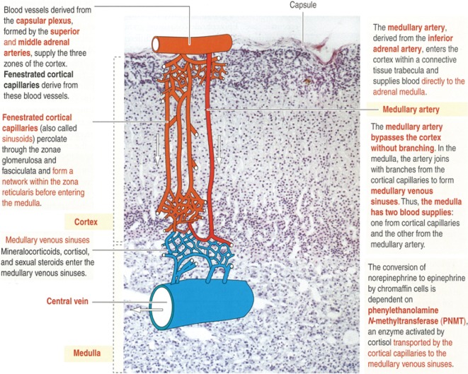
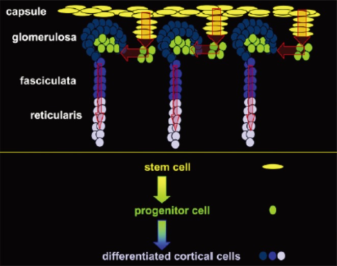
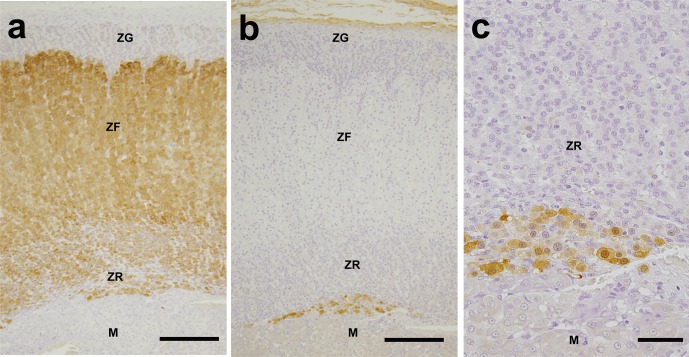
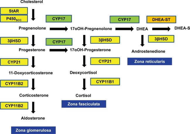
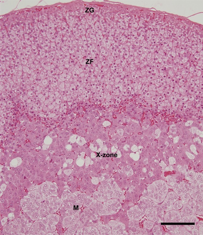
The adrenal gland has characteristic morphological and biochemical features that render it particularly susceptible to the actions of xenobiotics. As is the case with other endocrine organs, the adrenal gland is under the control of upstream organs (hypothalamic-pituitary system) in vivo, often making it difficult to elucidate the mode of toxicity of a test article. It is very important, especially for pharmaceuticals, to determine whether a test article-related change is caused by a direct effect or other associated factors. In addition, antemortem data, including clinical signs, body weight, food consumption and clinical pathology, and postmortem data, including gross pathology, organ weight and histopathologic examination of the adrenal glands and other related organs, should be carefully monitored and evaluated. During evaluation, the following should also be taken into account: (1) species, sex and age of animals used, (2) metabolic activation by a cytochrome P450 enzyme(s) and (3) physicochemical properties and the metabolic pathway of the test article. In this review, we describe the following crucial points for toxicologic pathologists to consider when evaluating adrenal toxicity: functional anatomy, blood supply, hormone production in each compartment, steroid biosynthesis, potential medulla-cortex interaction, and species and gender differences in anatomical features and other features of the adrenal gland which could affect vulnerability to toxic effects. Finally practical approaches for evaluating adrenal toxicity in nonclinical safety studies are discussed.

Keywords: adrenal cortex; adrenal medulla; nonclinical toxicity; species differences; steroidogenesis.


Figures

Fig. 1.
Fig. 1.
Cross sections of normal adrenal glands from a a) mouse, b) rat, c) dog and d) monkey. The adrenal glands are divided into two distinct endocrine tissues, the cortex and medulla. The cortex is characterized histologically by three layers, namely, the zona glomerulosa, zona fasciculata and zona reticularis. Note that the zona glomerulosa in the dog has a very different appearance compared with other species and consists of relatively large, flattened cells, which stain palely and are stacked in large loops (Fig. 1c). The zona reticularis is not clearly distinguishable in some rodents, particularly in the mouse. This zone is more distinct in rats compared with mice (Figs. 1a and b). ZG, zona glomerulosa; ZF, zona fasciculata; ZR, zona reticularis; M, medulla. HE stain. Bars = 100 µm (a, b), 200 µm (c, d).
Fig. 2.
Fig. 2.
Blood supply to the adrenal gland. Note that the dual blood supply to the medulla results in the transport of glucocorticoids necessary for PNMT activation and the supply of fresh blood to the medulla, which is required for rapid response to stress. These unique anatomical features of the adrenal blood supply are important for its function and conversely also contribute to the development of lesions. From Kierszenbaum AL with permission of Elsevier Ltd.
Fig. 3.
Fig. 3.
Hypothetical model of stem and progenitor cell centripetal migration and differentiation into steroidogenically competent adrenocortical cells. The stem or undifferentiated cell zone located in the innermost portion of the zona glomerulosa and the outermost portion of the zona fasciculata is the site for cell replication. This population may provide a pool of progenitors that differentiate into the neighboring zones. From Kim AC and Hammer GD with permission of Elsevier Ltd.
Fig. 4.
Fig. 4.
Cross sections of a normal adrenal gland from a monkey. Immunohistochemistry of (a) CYP17 and (b) low- and (c) high-magnification images of immunohistochemistry of DHEA-ST. The immunoreactivity of CYP17 is robust in the zona fasciculata and reticularis (Fig. 4a). The immunoreactivity of DHEA-ST is detected in the zona reticularis (Fig. 4b and c). ZG, zona glomerulosa; ZF, zona fasciculata; ZR, zona reticularis; M, medulla. Bars = 200 µm (a, b), 50 µm (c).
Fig. 5.
Fig. 5.
Pathways of adrenal steroid biosynthesis. StAR, steroid acute regulatory protein; P450SCC, P450 side chain cleavage enzyme; 3βHSD, 3β-hydroxysteroid dehydrogenase; DHEA, dehydroepiandrosterone; DHEA-ST, dehydroepiandrosterone sulfotransferase; DHEA-S, dehydroepiandrosterone sulphate.
Fig. 6.
Fig. 6.
X-zone in a cross section of the normal adrenal gland from a female mouse. The X-zone is a specific feature of the mouse adrenal cortex, a putative postpartal remnant of the fetal adrenal zone located at the junction of the cortex and medulla. Its precise function remains unknown, though it may be similar to the fetal zone in primates. ZG, zona glomerulosa; ZF, zona fasciculata; M, medulla. HE stain. Bar = 100 µm.

References

    1. Hammer GD, Parker KL, and Schimmer BP. Minireview: transcriptional regulation of adrenocortical development. Endocrinology. 146: 1018–1024. 2005. - PubMed
    1. Vinson GP, Whitehouse BJ, and Hinson JP. The Adrenal Cortex. Prentice Hall, New Jersey. 1992.
    1. Kierszenbaum AL. Endocrine system. In: Histology and Cell Biology: An Introduction to Pathology, 2nd ed. Elsevier Ltd., Philadelphia. 537–567. 2007.
    1. Hornsby PJ. The regulation of adrenocortical function by control of growth and structure. In: Adrenal Cortex. DC Anderson and JSD Winter (eds). Butterworth, London. 1–31. 1985.
    1. Hornsby PJ. Physiological and pathological effects of steroids on the function of the adrenal cortex. J Steroid Biochem. 27: 1161–1171. 1987. - PubMed