

doi: 10.1155/2015/942517.
Epub 2015 Sep 9.
Galactomannan Downregulates the Inflammation Responses in Human Macrophages via NFκB2/p100
Affiliations
- PMID: 26441484
- PMCID: PMC4579314
- DOI: 10.1155/2015/942517
Item in Clipboard
Galactomannan Downregulates the Inflammation Responses in Human Macrophages via NFκB2/p100
Mediators Inflamm.
2015.
Abstract
We show that galactomannan, a polysaccharide consisting of a mannose backbone with galactose side groups present on the cell wall of several fungi, induces a reprogramming of the inflammatory response in human macrophages through dectin-1 receptor. The nuclear factor kappa-light-chain-enhancer of activated B cells 2 (NFκB2)/p100 was overexpressed after galactomannan challenge. Knocking down NFκB2/p100 using small interfering RNA (siRNA) indicated that NFκB2/p100 expression is a crucial factor in the progression of the galactomannan-induced refractoriness. The data presented in this study could be used as a modulator of inflammatory response in clinical situations where refractory state is required.
Figures
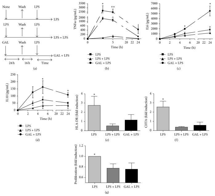
Galactomannan (GAL) attenuates the LPS-induced inflammation in human macrophages. (a) Schematic representation of the ET model used for this study. The cultures of human macrophages were or not pretreated with 10 ng/mL LPS or 10 μg/mL GAL for 24 h, washed twice with PBS, cultured in medium for 16 h, and restimulated with 10 ng/mL LPS for the indicated times. Supernatants were harvested at the indicated times (3 h, 5 h, and 24 h) and TNFα (b), IL6 (c), and IL10 (d) proteins levels were evaluated by CBA (n = 5); ∗
p < 0.05, ∗∗
p < 0.01 comparing GAL + LPS with LPS. (e) Cell surface expression of HLA-DR was analyzed by flow cytometry. (n = 5), ∗
p < 0.05, comparing GAL + LPS with LPS 24 h. (f) Total mRNA was isolated from cells, cDNA was synthesized and CIITA real-time quantitative PCR was performed. (n = 4), ∗
p < 0.05 comparing GAL + LPS with LPS 24 h. (g) Human heterologous lymphocytes were labeled with the membrane stain PKH2 Green Fluorescent Cell Linker Kit (Sigma-Aldrich) and cocultured with LPS, LPS + LPS, or GAL + LPS macrophages for 5 days. Lymphocyte proliferation was measured as a loss of green fluorescence intensity in the CD3+ gate, for this analysis. The fold induction is shown. ∗
p < 0.05, comparing GAL + LPS with LPS.
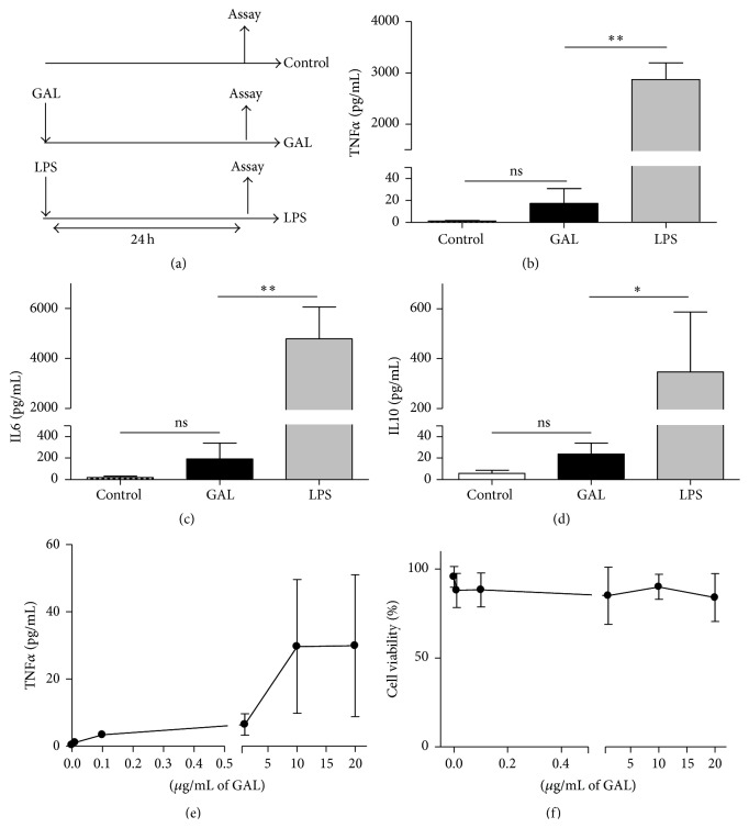
GAL is not able to induce an inflammatory response. (a) Schematic representation of the model used for this study. The cultures of human macrophages were treated with 10 ng/mL LPS or 10 μg/mL GAL for 24 h. The controls were not treated. Supernatants were harvested and TNFα (b), IL6 (c), and IL-10 (d) proteins levels were evaluated by CBA (n = 6); ∗
p < 0.05, ∗∗
p < 0.01 compared with LPS. (e) The culture of human macrophages was treated with GAL at various concentrations for 24 h. Supernatants were harvested and the TNFα protein levels were evaluated by CBA (n = 3). (f) The culture of human macrophages was treated with GAL at various concentrations for 24 h. Cells were harvested, and intracellular cells stained with 7AAD were analyzed by flow cytometry. The 7AAD negative cells are represented (percentage of cell viability, n = 4).
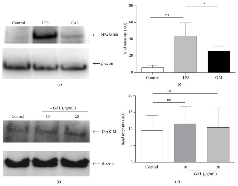
The NFκB2/p100 accumulation after the first LPS or GAL challenge. Cultures of human macrophages were challenged with 10 ng/mL LPS or 10 μg/mL GAL for 24 h. The controls were not treated. The levels of NFκB2/p100 or IRAK-M and β-actin were studied by Western blot analysis of the cytosolic fraction. (a) A NFκB2/p100 standard blot is shown (n = 6). (b) Densitometry analysis: arbitrary units [AU] of NFκB2/p100 bands are normalized with respect to β-actin (n = 6). ∗
p < 0.05, ∗∗
p < 0.01 compared with LPS. (c) A IRAK-M standard blot is shown (n = 3). (d) Densitometry analysis: arbitrary units [AU] of IRAK-M bands are normalized with respect to β-actin (n = 3).
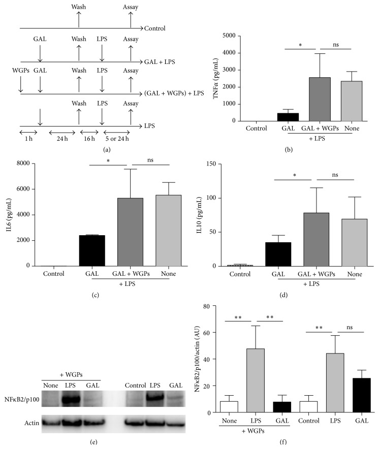
GAL is mediated by dectin-1 receptor. (a) Schematic representation of the inhibitor receptor of GAL model used for this study. The cultures of human macrophages were pretreated with or without WGPs (dectin-1 receptor inhibitor) 200 μg/mL for 1 hour. Next, GAL was added to the cells for 24 h, and then cells were washed three times and cultured in resting for 16 h. Cells were challenged with 10 ng/mL LPS for 5 h or 24 h. The controls were not pretreated. Supernatants were harvested at 5 h for TNFα (b), 24 h for IL6 (c), and 5 h for IL10 (d). Proteins levels were evaluated by CBA (n = 4); ∗
p < 0.05 compared with LPS. Levels of NFκB2/p100 and β-actin were studied by Western blot analysis of the cytosolic fraction. (e) A NFκB2/p100 standard blot is shown (n = 3). (f) Densitometry analysis: arbitrary units [AU] of NFκB2/p100 bands are normalized with respect to β-actin (n = 3). ∗∗
p < 0.01 compared with LPS.
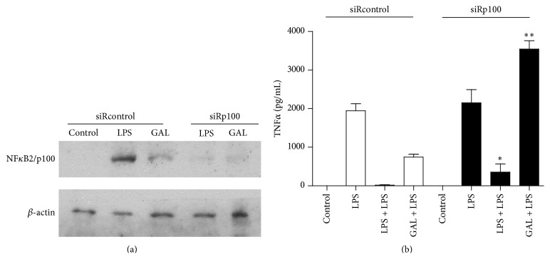
The attenuated inflammation effect induced by GAL is reverted by small interfering RNA of NFκB2/p100. Cultures of siRp100 and sirRcontrol transfected human monocyte/macrophages were treated with 10 g/mL GAL or 10 ng/mL LPS for 24 h. The controls were not treated. The levels of NFκB2/p100 and β-actin were studied by Western blot analysis of the cytosolic fraction. (a) A NFκB2/p100 and β-actin standard blot are shown (n = 5). (b) TNF protein level was evaluated by CBA (n = 4); ∗
p < 0.05, ∗∗
p < 0.01 compared with siRcontrol.
References
Publication types
MeSH terms
Substances
LinkOut - more resources
Full Text Sources
Other Literature Sources
