

Neurodegeneration and microtubule dynamics: death by a thousand cuts
- PMID: 26441521
- PMCID: PMC4563776
- DOI: 10.3389/fncel.2015.00343
Neurodegeneration and microtubule dynamics: death by a thousand cuts
Erratum in
-
Corrigendum: Neurodegeneration and microtubule dynamics: death by a thousand cuts.Front Cell Neurosci. 2016 Feb 9;10:26. doi: 10.3389/fncel.2016.00026. eCollection 2016. Front Cell Neurosci. 2016. PMID: 26903811 Free PMC article.
Abstract
Microtubules form important cytoskeletal structures that play a role in establishing and maintaining neuronal polarity, regulating neuronal morphology, transporting cargo, and scaffolding signaling molecules to form signaling hubs. Within a neuronal cell, microtubules are found to have variable lengths and can be both stable and dynamic. Microtubule associated proteins, post-translational modifications of tubulin subunits, microtubule severing enzymes, and signaling molecules are all known to influence both stable and dynamic pools of microtubules. Microtubule dynamics, the process of interconversion between stable and dynamic pools, and the proportions of these two pools have the potential to influence a wide variety of cellular processes. Reduced microtubule stability has been observed in several neurodegenerative diseases such as Alzheimer's disease (AD), Parkinson's disease (PD), Amyotrophic Lateral Sclerosis (ALS), and tauopathies like Progressive Supranuclear Palsy. Hyperstable microtubules, as seen in Hereditary Spastic Paraplegia (HSP), also lead to neurodegeneration. Therefore, the ratio of stable and dynamic microtubules is likely to be important for neuronal function and perturbation in microtubule dynamics might contribute to disease progression.
Keywords: Alzheimer's disease; Parkinson disease; dying back; hyperstable microtubules; microtuble stability; microtubule signaling hubs.
Figures
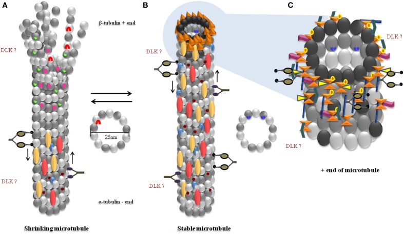
 ), β-Tubulin (
 ), GTP bound β-Tubulin (
 ), α/β heterodimer (
 ), Kinesin motor (
 ), Dynein motor (
 ). Present on less stable microtubules: Tyrosination (
 ), Gαs (
 ), LRRK2 (
 ). Present on stable microtubules: MAP1 (axon and dendrites) (
 ), TAU (axon) (
 ), MAP2 (
 ), Acetylation (
 ), Gβγ (
 ), +end binding proteins (
 ). Proteins present on the +end or fast growing end of microtubules (+TIPs): EBP1/2/3 (
 ), CLIP170 (
 ), CLASPS (
 ), APC (
 ), RHO GEF2 (
 ), MACF (
 ).
).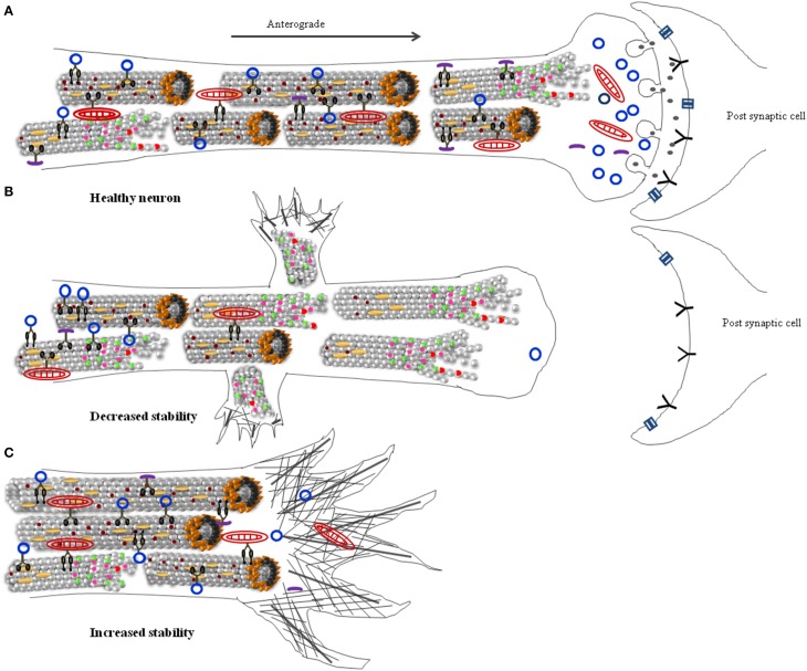
 ), depolymerzing microtubule (
 ), microtubule associated proteins (
 ), microtubule plus end binding proteins (
 ), mitochondria (
 ), membranous cargo (
 ), non-membranous cargo (
 ), Kinesin motor (
 ), Dynein motor (
 ), Actin filaments (
 ), Actin bundles (
 ), neurotransmitters (
 ), channels (
 ), neurotransmitter receptors (
 ).
).References
Publication types
LinkOut - more resources
Full Text Sources
Other Literature Sources
Miscellaneous