Review
. 2015 Sep 25:9:117.
doi: 10.3389/fnana.2015.00117. eCollection 2015.

Morphological changes of glutamatergic synapses in animal models of Parkinson's disease

Affiliations
Review

Morphological changes of glutamatergic synapses in animal models of Parkinson's disease

Rosa M Villalba et al. Front Neuroanat. .

Abstract

The striatum and the subthalamic nucleus (STN) are the main entry doors for extrinsic inputs to reach the basal ganglia (BG) circuitry. The cerebral cortex, thalamus and brainstem are the key sources of glutamatergic inputs to these nuclei. There is anatomical, functional and neurochemical evidence that glutamatergic neurotransmission is altered in the striatum and STN of animal models of Parkinson's disease (PD) and that these changes may contribute to aberrant network neuronal activity in the BG-thalamocortical circuitry. Postmortem studies of animal models and PD patients have revealed significant pathology of glutamatergic synapses, dendritic spines and microcircuits in the striatum of parkinsonians. More recent findings have also demonstrated a significant breakdown of the glutamatergic corticosubthalamic system in parkinsonian monkeys. In this review, we will discuss evidence for synaptic glutamatergic dysfunction and pathology of cortical and thalamic inputs to the striatum and STN in models of PD. The potential functional implication of these alterations on synaptic integration, processing and transmission of extrinsic information through the BG circuits will be considered. Finally, the significance of these pathological changes in the pathophysiology of motor and non-motor symptoms in PD will be examined.

Keywords: Parkinson’s disease; astrocytes plasticity; glutamatergic synapses; striatum; subthalamic nucleus; synaptic plasticity; vGluT.


Figures
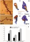
Figure 1
Figure 1
Dendritic spines in the monkey striatum. (A,B) Light micrographs of dendrites from Golgi-impregnated medium spiny neurons (MSNs) in the caudate nucleus of a control (A) and a 1-methyl-4-phenyl-1,2,3,6-tetrahydropyridine (MPTP)-treated (B) monkey. Note the dramatic spine loss of the dendrite of the MSN from the MPTP-treated monkey compared with control. (C1–D2) Three-dimension (3D)-reconstructed images of glutamatergic axo-spinous synapses from control (C1,C2) and MPTP-treated (D1,D2) monkeys. (E) Histogram comparing the morphometric measurements (mean ± SEM) for spine volume (μm3), post-synaptic density (PSD) area (μm2) and terminal volume (μm3) of structural elements at corticostriatal (vGluT1-positive) glutamatergic synapses using 3D reconstruction method of serial ultrathin sections collected from 30 axo-spinous synapses in each group from three control and three MPTP-treated animals. The spine volumes, the PSD areas, and the volume of vGluT1-containing terminals are significantly larger in MPTP-treated parkinsonian monkeys than in controls (*t-test; p < 0.001). Scale bar in (B) (applied to A) = 5 μm (See Villalba et al., ; Villalba and Smith, 2010, 2011a, 2013).
Figure 2
Figure 2
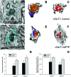
Tripartite synapses (TS) in the monkey striatum. (A,D) Electron micrographs of perisynaptic astrocytic processes (Ast) wrapping a vGluT1 axo-spinous synapse in control (A) and MPTP-treated (D) animal. (B,C,E,F) The three-dimensional (3D) reconstruction of a vGluT1-immunoreactive TS highlight the differences in the extensions of the astrocytic processes between control (B,C) and MPTP (E,F). In the TS of control animals, the perimeters of the axon-spinous interfaces were only partially surrounded by astroglial processes (B,C). In MPTP-treated animals, TS vGluT1-containing synapses displayed a large increase in astroglial processes ensheatment (E,F). (G) Histograms comparing the surface area of perisynaptic glia associated with vGluT1- and vGluT2-immunopositive axo-spinous synapses in control (N = 3) and MPTP-treated (N = 3) monkeys (mean ± SEM). The surface of the perisynaptic glia was significant larger (*, t-test, p = 0.017 for vGluT1 and p = 0.006 for vGluT2) in MPTP-parkinsonian monkeys than in control. (H) Histograms comparing the ratio of the volume of the perisynaptic glia over the total volume of spine and the axon terminals in TS formed by vGluT1- or vGluT2-immunoreactive terminals. This ratio was significantly larger in MPTP than in control condition (*, t-test, p = 0.049 for vGluT1 and p = 0.028 for vGluT2). No significant difference was found between TS formed by vGluT1- or vGluT2-immunoreactive terminals. Total number of reconstructed spines = 32 (8 per group). Statistics were performed by using SigmaPlot (version 11.0). Abbreviations: Ast, astrocyte; PSD, post-synaptic density; Sp, dendritic spine; T, axon terminal (see Villalba and Smith, for details).
Figure 3
Figure 3
vGluT1-positive innervation in the monkey subthalamic nucleus (STN). (A) Light micrograph showing vGluT1-positive varicose processes. (B) Average density (mean ± SEM; N = 3) of vGluT1-immunoreactive varicosities in the dorsolateral STN of normal and parkinsonian monkeys (*, t-test, p = 0.012). (C) Comparison of the average STN volume (mean ± SEM; N = 3) between normal and parkinsonian monkeys. (D) Electron micrograph showing an asymmetric synapse (arrows) in the dorsolateral monkey STN. (E) Average density (mean ± SEM; N = 3) of vGluT1-immunopositive terminals in the dorsolateral STN of normal and parkinsonian monkeys (*, t-test, p = 0.02). (F) Average density (mean ± SEM; N = 3) of asymmetric synapses in the dorsolateral STN of normal and pakinsonian monkeys (*, t-test, p = 0.029). (G,H) Electron micrographs showing vGluT1-containing terminals forming asymmetric synapses with a spine (G) and a dendritic shaft (H). (I) Post-synaptic targets of vGluT1-immunopositive terminals in the dorsolateral STN. No differences were found in the proportion of vGluT1-immunoreactive terminals forming asymmetric synapses with dendritic shafts and spines in normal and parkinsonian animals. Scale bar A = 10 μm and in (D; applies also to G) and H = 0.2 μm. Abbreviations: Den, dendrite; Sp, dendritic spine; T, axon terminal (See Mathai et al., 2015).
Figure 4
Figure 4
Schematic showing morphological changes in dendritic spines and glutamatergic afferents in striatal MSNs and projection neurons in the STN in MPTP-treated parkinsonian monkeys. In the striatum of parkinsonian monkeys, there is a significant reduction in the density of Sp on MSNs, but the remaining spines and terminals display an increase in volume. The size of the PSD at corticostriatal and thalamostriatal synapses is also increased and more commonly perforated in parkinsonian animals than controls. In the STN, there is an overall decrease in the prevalence of vGluT1-positive cortical terminals in contact with dendrites and spines of STN neurons in parkinsonian animals. Potential changes in the ultrastructure of spines and afferent glutamatergic terminals, as shown in the striatum, remain to be determined in the STN.

References

    1. Alexander G. E., DeLong M. R., Strick P. L. (1986). Parallel organization of functionally segregated circuits linking basal ganglia and cortex. Annu. Rev. Neurosci. 9, 357–381. 10.1146/annurev.neuro.9.1.357 - DOI - PubMed
    1. Antal M., Beneduce B. M., Regehr W. G. (2014). The substantia nigra conveys target-dependent excitatory and inhibitory outputs from the basal ganglia to the thalamus. J. Neurosci. 34, 8032–8042. 10.1523/JNEUROSCI.0236-14.2014 - DOI - PMC - PubMed
    1. Arbuthnott G. W., Wickens J. (2007). Space, time and dopamine. Trends Neurosci. 30, 62–69. 10.1016/j.tins.2006.12.003 - DOI - PubMed
    1. Arbuthnott G. W., Ingham C. A., Wickens J. R. (2000). Dopamine and synaptic plasticity in the neostriatum. J. Anat. 196, 587–596. 10.1046/j.1469-7580.2000.19640587.x - DOI - PMC - PubMed
    1. Aymerich M. S., Barroso-Chinea P., Pérez-Manso M., Muñoz-Patiño A. M., Moreno-Igoa M., González-Hernández T., et al. . (2006). Consequences of unilateral nigrostriatal denervation on the thalamostriatal pathway in rats. Eur. J. Neurosci. 23, 2099–2108. 10.1111/j.1460-9568.2006.04741.x - DOI - PubMed

LinkOut - more resources